Supporting book: Robbins “Basic Pathology” (10th edition)
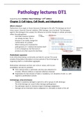
Chapter 2: Cell Injury, Cell Death, and Adaptations
What is disease?
Dysfunction of an organ or tissue, because of damage to the cells. The damage can be of
many causes: chemical, thermal, radiation, DNA damage, micro bacterial. The damaging
agent is the etiology(or the causes), the influence on and the changes in cellular processes
reflect the pathogenesis.
- One little missense mutation
can already be fatal. This is
because it can totally change the
conformation of a protein.
- When looking at for example the
pathogenesis of V. cholerae the bacteria itself
is not so dangerous, but the toxins it
produces are what causes the diarrhoea
Multicellular organisms
A lot of the times being a community can help for survival. The social
amoeba Dictyostelium discoideum survives periods of food shortage by
organising itself in a multicellular aggregate.
Multicellular individuals are their own niche in nature, with its own
possibilities, but also its problems.
➔ Their internal milieu is optimised, and thus also attractive for
intruders. Effective defence is required (infectious diseases)
➔ Organisation & clear division of tasks is mandatory, incl. discipline of cells, i.e., with
regards to proliferation (cancer)
Cell damage, stress & stressors
- Disease is caused by damage to (part of) a cell or group of cells (etiology)
- The initial damage can cause further damage (pathogenesis)
- The cell/organ reacts to minimize impact of damage (adaptation)
- Damage can be reversible, lead to adaptation or, ultimately to death of the cell
1
,When cells are under stress, they can do different
things. To illustrate this, let’s look at the reaction of
myocardial cells; when there is an increase load of
blood, myocardial cells can adapt by thickening the
wall (Hypertrophy). When the blood flow is decreased
(by an infarction for example), ischemia will lead to cell
injury, that in turn leads to cell death.
There are more ways cells & tissues can adapt
(reversible injury):
1. Hypertrophy
This is what happens in myocytes after an increased load. Remember that with hypertrophy
there is an increase in the size of cells, NO increase in the number of cells. When the stress is
not relieved after hypertrophy, significant cell injury can still happen.
The mechanisms driving hypertrophy involve:
- Mechanical triggers; like stretch
- Soluble mediators; stimulate cell growth,
such as growth factors and adrenergic
hormones.
The stimuli will indirectly induce more genes,
which in turn stimulate synthesis of many cellular
proteins and in turn cause hypertrophy. The
myocardial fibres can enlarge to a certain limit.
Surpassing this limit eventually results in
ventricular dilation and ultimately cardiac failure.
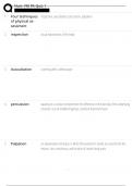
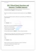
2. Hyperplasia
This is the contrary of hypertrophy. Instead of an increase in the size of cells, the number of
cells increase when hyperplasia is induced. It will only take place in populations that are
capable of replication. Sometimes it can occur concurrently with hypertrophy and often in
response to the same stimuli.
There are two types of hyperplasia;
- Hormonal hyperplasia; this is illustrated in
the picture right. When a woman is in
puberty or pregnant, the glandular
epithelium of the female breast will
proliferate.
- Compensatory hyperplasia; this is when residual
tissue grows after removal or loss of part of an organ. For example, when part of a
liver is resected, mitotic activity in the remaining cells begins restoring the liver to its
normal size.
In pathologic hyperplasia, the excessive hormonal or growth factor stimulation are the major
cause. When a woman has endometrial hyperplasia, there is a disturbance in the balance of
epithelial proliferation after menstruation.
2
,Hyperplasia is not the same as cancer, because the hyperplastic process remains controlled. If
the signals that initiate it abate, the hyperplasia disappears. In cancer the cell proliferation is
uncontrolled. But(!), hyperplasia can constitute a fertile soil in which cancers may eventually
arise.
3. Atrophy
This is a decrease of tissue by decrease of cell size/number. These atrophic cells are not
dead. Causes of atrophy include
- A decreased workload (e.g., immobilization of a limb to permit healing of a fracture)
- Loss of innervation
- Diminished blood supply
- Inadequate nutrition
- Loss of endocrine stimulation
- Aging
The changes are similar in pathological situations and physiologic situations. Cellular atrophy
results from a combination of decreased protein synthesis and increased protein
degradation.
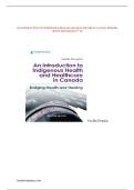
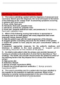
The picture right depicts the difference between a normal brain (A) and a
82-year-old brain (B) with atherosclerotic disease. This state of atrophy is
caused by aging and reduced blood supply.
In many situations, atrophy is associated with autophagy, with resulting
increases in the number of autophagic vacuoles. In other situations, atrophy is Autophagy is the
associated with apoptosis and proteasomal degradation. process in which the
starved cell eats its own
4. Metaplasia organelles in an attempt
This is the replacement of one tissue by a (normal) other tissue. Metaplasia is to survive.
thought to arise by the reprogramming of stem cells to differentiate along a
new pathway rather than a phenotypic change of already differentiated cells.
When you smoke, all these toxins are entering the
airways. The epithelial doesn’t like this so its tissue
will be replaced by means of metaplasia. One
disadvantage in this cell replacement is that we don’t
know how this tissue is going to behave. So, it can
also be cancerous cells that will proliferate. A lot of
the times the epithelium will turn into squamous
epithelium. This may be able to survive the chemicals
in cigarette smoke that the more fragile specialized
epithelium would not tolerate. Although its survival
advantages, important protective mechanisms are lost, such as mucus secretion and ciliary
clearance of particulate matter.
3
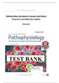
,Left, you see normal ciliated
bronchial epithelium and right you
see squamous metaplastic
bronchial epithelium. This does not
contain the cilia that seem like
actual hairs.
Next to adaptation, there are also different ways cells can be injured what will lead to
cell death:
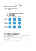
Oxygen shortage
Exemplified, oxygen is very important in kidney cells. If you don’t have enough oxygen in
your kidney cells, then sodium pumps will die, and the cell too. This because the water
balance cannot be maintained, and therefore, the cell will swell and explode.
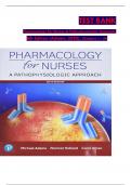
A: normal kidney tubules with viable epithelial
cells.
B: early (reversible) ischemic injury showing
surface blebs, increased eosinophilia of
cytoplasm, and swelling of occasional cells.
C: Necrotic (irreversible) injury of epithelial
cells, with loss of nuclei and fragmentation of
cells and leakage of contents.
Necrosis
This is a form of cell death in which cellular membranes fall apart, and cellular
enzymes leak out an ultimately digest the cell.
The biochemical mechanisms of necrosis vary with different injurious stimuli.
These mechanisms include:
- Failure of energy generation in the form of ATP because of reduced
oxygen supply or mitochondrial damage
- Damage to cellular membranes, including the plasma membrane and
lysosomal membranes, which results in leakage of cellular contents
including enzymes
- Irreversible damage to cellular lipids, proteins and nucleic acids, which
may be caused by reactive oxygen species (ROS)
4
, The most important thing to remember of necrosis, is
that it is pathological and it produces inflammation.
Types of necrosis:
o Coagulative necrosis: The underlying tissue
architecture is preserved for at least several
days after death of cells in the tissue.
o Liquefactive necrosis: Dead cells are completely digested by leukocytes,
transforming the tissue into a viscous liquid that is eventually removed by
phagocytes (pus).
o Gangrenous necrosis: For example, when a limb has lost its blood supply
it can undergo coagulative necrosis involving multiple tissue layers.
o Caseous necrosis: (seen in tuberculosis) It means ‘cheese like’, referring
to the friable yellow-white appearance of the area. Caseous necrosis is
often surrounded by a collection of macrophages and other inflammatory
cells.
o Fat necrosis: Typically a result from the release of activated pancreatic
lipases into the substance of the pancreas and the peritoneal cavity.
Released fatty acids combine with calcium to produce grossly visible
chalky white areas (fat saponification)
o Fibrinoid necrosis: Special form. It usually occurs in immune reactions
in which complexes of antigens and antibodies are deposited in the
walls of blood vessels, but it also may occur in severe hypertension.
Apoptosis
https://www.youtube.com/watch?v=SyvOPXeg4ig
This is a pathway of cell death in which cells activate enzymes that degrade
the cells’ own nuclear DNA and nuclear and cytoplasmic proteins.
➔ Programmed cell death
When apoptosis?
- Embryonal development
- Normal tissue homeostasis
- Selection (of early maturational stages of lymphocytes by antigen receptors)
- Involution or atrophy
- Termination of inflammatory response or immune reaction
- Elimination of virus-infected cells or cells with (oncogenic and other) mutations, by
CTL
- Elimination of stressed cells by NK cell
- Elimination of damaged cells
5
Chapter 2: Cell Injury, Cell Death, and Adaptations
What is disease?
Dysfunction of an organ or tissue, because of damage to the cells. The damage can be of
many causes: chemical, thermal, radiation, DNA damage, micro bacterial. The damaging
agent is the etiology(or the causes), the influence on and the changes in cellular processes
reflect the pathogenesis.
- One little missense mutation
can already be fatal. This is
because it can totally change the
conformation of a protein.
- When looking at for example the
pathogenesis of V. cholerae the bacteria itself
is not so dangerous, but the toxins it
produces are what causes the diarrhoea
Multicellular organisms
A lot of the times being a community can help for survival. The social
amoeba Dictyostelium discoideum survives periods of food shortage by
organising itself in a multicellular aggregate.
Multicellular individuals are their own niche in nature, with its own
possibilities, but also its problems.
➔ Their internal milieu is optimised, and thus also attractive for
intruders. Effective defence is required (infectious diseases)
➔ Organisation & clear division of tasks is mandatory, incl. discipline of cells, i.e., with
regards to proliferation (cancer)
Cell damage, stress & stressors
- Disease is caused by damage to (part of) a cell or group of cells (etiology)
- The initial damage can cause further damage (pathogenesis)
- The cell/organ reacts to minimize impact of damage (adaptation)
- Damage can be reversible, lead to adaptation or, ultimately to death of the cell
1
,When cells are under stress, they can do different
things. To illustrate this, let’s look at the reaction of
myocardial cells; when there is an increase load of
blood, myocardial cells can adapt by thickening the
wall (Hypertrophy). When the blood flow is decreased
(by an infarction for example), ischemia will lead to cell
injury, that in turn leads to cell death.
There are more ways cells & tissues can adapt
(reversible injury):
1. Hypertrophy
This is what happens in myocytes after an increased load. Remember that with hypertrophy
there is an increase in the size of cells, NO increase in the number of cells. When the stress is
not relieved after hypertrophy, significant cell injury can still happen.
The mechanisms driving hypertrophy involve:
- Mechanical triggers; like stretch
- Soluble mediators; stimulate cell growth,
such as growth factors and adrenergic
hormones.
The stimuli will indirectly induce more genes,
which in turn stimulate synthesis of many cellular
proteins and in turn cause hypertrophy. The
myocardial fibres can enlarge to a certain limit.
Surpassing this limit eventually results in
ventricular dilation and ultimately cardiac failure.
2. Hyperplasia
This is the contrary of hypertrophy. Instead of an increase in the size of cells, the number of
cells increase when hyperplasia is induced. It will only take place in populations that are
capable of replication. Sometimes it can occur concurrently with hypertrophy and often in
response to the same stimuli.
There are two types of hyperplasia;
- Hormonal hyperplasia; this is illustrated in
the picture right. When a woman is in
puberty or pregnant, the glandular
epithelium of the female breast will
proliferate.
- Compensatory hyperplasia; this is when residual
tissue grows after removal or loss of part of an organ. For example, when part of a
liver is resected, mitotic activity in the remaining cells begins restoring the liver to its
normal size.
In pathologic hyperplasia, the excessive hormonal or growth factor stimulation are the major
cause. When a woman has endometrial hyperplasia, there is a disturbance in the balance of
epithelial proliferation after menstruation.
2
,Hyperplasia is not the same as cancer, because the hyperplastic process remains controlled. If
the signals that initiate it abate, the hyperplasia disappears. In cancer the cell proliferation is
uncontrolled. But(!), hyperplasia can constitute a fertile soil in which cancers may eventually
arise.
3. Atrophy
This is a decrease of tissue by decrease of cell size/number. These atrophic cells are not
dead. Causes of atrophy include
- A decreased workload (e.g., immobilization of a limb to permit healing of a fracture)
- Loss of innervation
- Diminished blood supply
- Inadequate nutrition
- Loss of endocrine stimulation
- Aging
The changes are similar in pathological situations and physiologic situations. Cellular atrophy
results from a combination of decreased protein synthesis and increased protein
degradation.
The picture right depicts the difference between a normal brain (A) and a
82-year-old brain (B) with atherosclerotic disease. This state of atrophy is
caused by aging and reduced blood supply.
In many situations, atrophy is associated with autophagy, with resulting
increases in the number of autophagic vacuoles. In other situations, atrophy is Autophagy is the
associated with apoptosis and proteasomal degradation. process in which the
starved cell eats its own
4. Metaplasia organelles in an attempt
This is the replacement of one tissue by a (normal) other tissue. Metaplasia is to survive.
thought to arise by the reprogramming of stem cells to differentiate along a
new pathway rather than a phenotypic change of already differentiated cells.
When you smoke, all these toxins are entering the
airways. The epithelial doesn’t like this so its tissue
will be replaced by means of metaplasia. One
disadvantage in this cell replacement is that we don’t
know how this tissue is going to behave. So, it can
also be cancerous cells that will proliferate. A lot of
the times the epithelium will turn into squamous
epithelium. This may be able to survive the chemicals
in cigarette smoke that the more fragile specialized
epithelium would not tolerate. Although its survival
advantages, important protective mechanisms are lost, such as mucus secretion and ciliary
clearance of particulate matter.
3
,Left, you see normal ciliated
bronchial epithelium and right you
see squamous metaplastic
bronchial epithelium. This does not
contain the cilia that seem like
actual hairs.
Next to adaptation, there are also different ways cells can be injured what will lead to
cell death:
Oxygen shortage
Exemplified, oxygen is very important in kidney cells. If you don’t have enough oxygen in
your kidney cells, then sodium pumps will die, and the cell too. This because the water
balance cannot be maintained, and therefore, the cell will swell and explode.
A: normal kidney tubules with viable epithelial
cells.
B: early (reversible) ischemic injury showing
surface blebs, increased eosinophilia of
cytoplasm, and swelling of occasional cells.
C: Necrotic (irreversible) injury of epithelial
cells, with loss of nuclei and fragmentation of
cells and leakage of contents.
Necrosis
This is a form of cell death in which cellular membranes fall apart, and cellular
enzymes leak out an ultimately digest the cell.
The biochemical mechanisms of necrosis vary with different injurious stimuli.
These mechanisms include:
- Failure of energy generation in the form of ATP because of reduced
oxygen supply or mitochondrial damage
- Damage to cellular membranes, including the plasma membrane and
lysosomal membranes, which results in leakage of cellular contents
including enzymes
- Irreversible damage to cellular lipids, proteins and nucleic acids, which
may be caused by reactive oxygen species (ROS)
4
, The most important thing to remember of necrosis, is
that it is pathological and it produces inflammation.
Types of necrosis:
o Coagulative necrosis: The underlying tissue
architecture is preserved for at least several
days after death of cells in the tissue.
o Liquefactive necrosis: Dead cells are completely digested by leukocytes,
transforming the tissue into a viscous liquid that is eventually removed by
phagocytes (pus).
o Gangrenous necrosis: For example, when a limb has lost its blood supply
it can undergo coagulative necrosis involving multiple tissue layers.
o Caseous necrosis: (seen in tuberculosis) It means ‘cheese like’, referring
to the friable yellow-white appearance of the area. Caseous necrosis is
often surrounded by a collection of macrophages and other inflammatory
cells.
o Fat necrosis: Typically a result from the release of activated pancreatic
lipases into the substance of the pancreas and the peritoneal cavity.
Released fatty acids combine with calcium to produce grossly visible
chalky white areas (fat saponification)
o Fibrinoid necrosis: Special form. It usually occurs in immune reactions
in which complexes of antigens and antibodies are deposited in the
walls of blood vessels, but it also may occur in severe hypertension.
Apoptosis
https://www.youtube.com/watch?v=SyvOPXeg4ig
This is a pathway of cell death in which cells activate enzymes that degrade
the cells’ own nuclear DNA and nuclear and cytoplasmic proteins.
➔ Programmed cell death
When apoptosis?
- Embryonal development
- Normal tissue homeostasis
- Selection (of early maturational stages of lymphocytes by antigen receptors)
- Involution or atrophy
- Termination of inflammatory response or immune reaction
- Elimination of virus-infected cells or cells with (oncogenic and other) mutations, by
CTL
- Elimination of stressed cells by NK cell
- Elimination of damaged cells
5