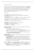
bMETHODS IN NEUROSCIENCE
Week 1 – Anatomy and connectomics 2
Week 2 – Optogenetics 14
Week 3 – Optogenetics II 22
Week 4 – Behavioral Testing 27
Week 5 – Electrophysiology methods 39
Week 6 – Genetic methods for systems neuroscience 49
Week 7 – Multi-photon imaging 57
,Week 1 – Anatomy and connectomics
The goal of anatomy
1) Describe the structural components of nervous system
a) Structure of receptors (e.g. AMPA receptor)
b) e.g. morphology of neurons (pyramidal neuron, stellate cell, etc.)
c) Organization of brain into regions (mapping)
2) Determine the connections between neurons, or between areas (the connectome)
3) Determine how structure changes over time (development, plasticity)
History
Technology (in order)
● 19th century: advances light microscopy → fixation tissue → staining
● 20th century: electron microscopy → tracing techniques → viral techniques →
genetic engineering
● 21st century: AI-assisted connectomics
Findings (in order)
● 19th century: neurons as individual cells → dendrites, axons, soma, information flow
→ demarcation brain areas
● Between 20th and 21st: synapse, vesicles, receptor structure → connection matrices,
cortical hierarchy → connections in microcircuit → full connectomes of simple
organisms
Learning goals exam:
● The light microscopy (19th century) came before electron microscopy (20th century)
● Advances in microscopy and the invention of the Golgi stain. Visualizing the
morphology and architecture of neurons and their circuits throughout the brain
● EM is better for analysis of connectome since it has nanometer resolution
History of the neuron: at the end of the 19th century, the concept of a “neuron” emerged as
a physically separate entity.
● At that time, mainstream theory was that the brain was a continuous single network,
they believed that there were no individual cells → reticular theory
○ The mind should be continuous and holistic
○ They knew there were cells in bodies, but did not think that the brain
consisted of separate ones.
○ Reticularists vs Neuronists:
● Reticularists: all neurons are connected in a continuous structure network
○ Otto F K Deiters
○ J. von Gerlach
○ C. Golgi: Nobel Prize in Physiology or Medicine, 1906
2
, ● Neuronists: cells are individuals elements that are separated from each other, no
continuous structure, and communicate via contact
○ Wilhelm His
○ Auguste Forel
○ S. Ramon y Cajal: Nobel Prize in Physiology or Medicine, 1906
● The debate was resolved by the development of the EM → provides proof that the
nervous system is composed of individual, discrete neurons that communicate with
each other via synapses
Fixation tissue
● Fixation: process of using chemical methods to preserve, stabilize, and strengthen a
biological specimen for subsequent histological procedures and microscopic analysis.
○ terminating ongoing biological reactions by “fixing” proteins into place
● Formaldehyde, CH2O (mid 19th century)
○ fixes proteins by forming methylene (CH2) bridges among amino acid
○ good for histological analysis, electron microscopy, tracing experiments,
fluorescence
○ not food for RNA and DNA because it disrupts the structure
● Immersion: placing small brains, or even entire animals, in fixative solutions
○ slow and efficient diffusion through tissue, Distance = K x √Time
○ Diffusion is random and takes much longer; every formaldehyde molecule has
to reach the inside of the brain, but since it is just diffusing randomly, you end
up only staining the outside of the brain, and do not reach the inside → takes
proportionally longer
○ Easier in small animals
● Perfusion: uses vascular system to deliver a fixative
○ Syringe needle into the left ventricle of the animal that is connected to the
perfusion pump → the pump pumps the fixative formaldehyde solution
through the entire body → reaches the nervous system
○ Thorough and quick
○ Adequately fixes all cells in the brain if properly performed
○ More difficult in smaller animals
Golgi-fill
● Golgi-stain: technique used to completely label individuals neurons + their processes
○ technological breakthrough end of 19th century
○ 1) Fixation of tissue
○ 2) Chemical reaction with silver nitrate (AgNO3)
■ Golgi-fill: when the entire neuron is filled with black
○ 3) Cut tissue into small sections for visualization
○ 4) Visualization with light microscopy
3
, ● Advantages:
○ Fill of the entire neuron, allows to see dendrites and axons
○ Sparse labelling of few neurons → good for visualization as only 5-10% of the
total number of cells are labeled → individual neurons stand out in a
background of numerous unlabeled cells
● Invented by Golgi, allowed visualization of individual neurons and
their connections
Visualization
● Many advances in late 19th century from light microscopy (Zeiss,
Abbe)
● Allowed to see dendrites, cell bodies, axons in details
● Synapses are too small for light microscopy
● Single dendrite and neurons are visible with light microscopy
● DNA is difficult is impossible to see with light microscopy
● Mouse brain is visible with the naked eye
Neuron doctrine
● Ramon y Cajal showed nervous system is made up of discrete cells
● Cajal’s Neuron doctrine
○ Flow of information from dendrites → soma → axon
○ Propagation of electric currents in this direction
○ Cajal improved the staining technique, and was able to
observe that nerve cells are discrete, individual units, rather
than a continuous network → neuron doctrine
● Hypothesis: neurons are the basic structural and functional units of the NS
● Revelation of morphological diversity of neurons and neural circuits in the brain
● Cajal was not able to directly observe synapses, due to the limits of light microscopy
of that time. It lacked the resolution to distinguish the small gap between neurons.
Nissl stain: basophilic (“base + loving”) stain used to visualize cell bodies
● Good for staining acidic molecules
● Labels negatively charged molecules like RNA and DNA
● Provides contrast to ribosomes & rough ER, enriched in neurons
● Visualizes the cell bodies, commonly used for brain atlases
○ Allows to see densities, so you can distinguish between
different layers
● Examples: differentiation of different cortical layers based on cell density, or
differentiation of areas (e.g. V1 and V2)
● What does it visualize: nuclei, cell bodies, useful for making brain atlases, you can
see the densities, tell apart that there are different layers
4