Cardiac Contraction and Inotropic effects
Key Points:
Describe mechanisms which increase and decrease [Ca2+]i
How changes in [Ca2+]i relate to contraction
Explain mechanisms by which drugs can increase cardiac contractility
Rise in [Ca2+]i – Central to Contraction
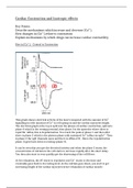
This graph shows electrical activity of the heart compared with the amount of Ca 2+
signalling in cells (amount of Ca2+ in cell) going on and the cardiac myocytes length.
The electrical graph at the top is split into the phases of cardiac contraction, split into
phase 4 which is the resting potential, then phase 0 is the upstroke where there is
rapid Na+ influx, this is depolarisation. You reach the peak at phase 1 and then after
there is phase 2 which is the plateau phase with sustained Ca 2+ influx via vgCa2+. Then
in phase 3 the vgK+ channels open and there is efflux of K+, this is the repolarization
phase. It gets back down to resting (phase 4).
It can be seen that you get the electrical activity and when the phase 2 starts, the
concentration of calcium in the cell starts to increase rapidly after the short delay.
You then also start to very quickly get the shortening of the cell fibres.
As for relaxation, the AP starts to repolarise and Ca 2+ starts to decrease and
eventually goes back to its resting level. As the calcium goes down, you start to get
increasing length of the cardiac myocytes hence relaxation of cardiac muscle.
, Action potential duration is around 200-500 miliseconds, the force of contraction is
proportional to [Ca2+]i.
During normal systole [Ca2+]i may be around 1mM, during exercise or something
similar you will be producing maximum contraction of your heart myocytes and the
[Ca2+]i is going to be around 10mM in order to produce this contraction (because
remember force of contraction is proportional to calcium concentration).
When resting i.e. normally, cell shortening in our heart is sub-maximal (i.e. not
maximum shortening, hence not max contraction) and this is because our calcium
levels are not at maximum level.
This is important because in skeletal muscle you just get the maximum release of
calcium into a fibre and get full contraction, skeletal muscle is regulated by fibre
recruitment. In the heart we have enough muscle fibres, we instead regulate the
force of contraction through calcium levels in the cells.
If the heart didn’t work this way, you would have maximum contraction all the time,
you wouldn’t be able to up-regulate or down-regulate your stroke volume.
How does electrical excitability contract cardiac myocytes?
Contraction is determined by an increase in intracellular Ca2+ levels, the higher the
increase in Ca2+, the greater the force of contraction.
Intracellular Ca2+ levels increase from 0.1mM to about 10mM.
Below we can see a diagram of what happens in an atrial/ventricular myocyte
When we get to phase 2 of the cardiac electrical activity, we have our influx of Ca 2+
down vgCa2+ (L type) which is sustained, they are relatively slow to start and slow to
turn off, which is why it is sustained. Eventually we start getting the efflux of K + as
vgCa2+ start to turn off.
So looking at diagram, we have phase 0 before this and the membrane gets
depolarised this causes the VDCC open up, Ca2+ influxes into the cell down its very
steep concentration gradient (Ca2+ is very low in the cell). This increases intracellular
Ca2+ through calcium induced calcium release.
Key Points:
Describe mechanisms which increase and decrease [Ca2+]i
How changes in [Ca2+]i relate to contraction
Explain mechanisms by which drugs can increase cardiac contractility
Rise in [Ca2+]i – Central to Contraction
This graph shows electrical activity of the heart compared with the amount of Ca 2+
signalling in cells (amount of Ca2+ in cell) going on and the cardiac myocytes length.
The electrical graph at the top is split into the phases of cardiac contraction, split into
phase 4 which is the resting potential, then phase 0 is the upstroke where there is
rapid Na+ influx, this is depolarisation. You reach the peak at phase 1 and then after
there is phase 2 which is the plateau phase with sustained Ca 2+ influx via vgCa2+. Then
in phase 3 the vgK+ channels open and there is efflux of K+, this is the repolarization
phase. It gets back down to resting (phase 4).
It can be seen that you get the electrical activity and when the phase 2 starts, the
concentration of calcium in the cell starts to increase rapidly after the short delay.
You then also start to very quickly get the shortening of the cell fibres.
As for relaxation, the AP starts to repolarise and Ca 2+ starts to decrease and
eventually goes back to its resting level. As the calcium goes down, you start to get
increasing length of the cardiac myocytes hence relaxation of cardiac muscle.
, Action potential duration is around 200-500 miliseconds, the force of contraction is
proportional to [Ca2+]i.
During normal systole [Ca2+]i may be around 1mM, during exercise or something
similar you will be producing maximum contraction of your heart myocytes and the
[Ca2+]i is going to be around 10mM in order to produce this contraction (because
remember force of contraction is proportional to calcium concentration).
When resting i.e. normally, cell shortening in our heart is sub-maximal (i.e. not
maximum shortening, hence not max contraction) and this is because our calcium
levels are not at maximum level.
This is important because in skeletal muscle you just get the maximum release of
calcium into a fibre and get full contraction, skeletal muscle is regulated by fibre
recruitment. In the heart we have enough muscle fibres, we instead regulate the
force of contraction through calcium levels in the cells.
If the heart didn’t work this way, you would have maximum contraction all the time,
you wouldn’t be able to up-regulate or down-regulate your stroke volume.
How does electrical excitability contract cardiac myocytes?
Contraction is determined by an increase in intracellular Ca2+ levels, the higher the
increase in Ca2+, the greater the force of contraction.
Intracellular Ca2+ levels increase from 0.1mM to about 10mM.
Below we can see a diagram of what happens in an atrial/ventricular myocyte
When we get to phase 2 of the cardiac electrical activity, we have our influx of Ca 2+
down vgCa2+ (L type) which is sustained, they are relatively slow to start and slow to
turn off, which is why it is sustained. Eventually we start getting the efflux of K + as
vgCa2+ start to turn off.
So looking at diagram, we have phase 0 before this and the membrane gets
depolarised this causes the VDCC open up, Ca2+ influxes into the cell down its very
steep concentration gradient (Ca2+ is very low in the cell). This increases intracellular
Ca2+ through calcium induced calcium release.