1
Innovative Therapeutics
1. Structure and stability of proteins (1-9-2025, Trombetta Lima) 2
2. Production of recombinant proteins (2-9-2025, Trombetta Lima) 5
3. Molecular biotechnology and genomics (3-9-2025, Wen Wu) 8
4. Biotherapeutics (4-9-2025, Sousa) 12
5. Development of RNA-based vaccines (5-9-2025, Sousa) 14
6. Molecular basis for the use of light in medicine (8-9-2025, Szymanski) 17
7. Biosimilars and ATMP’s (9-9-2025, Kosterink) 19
8. Immunogeniciteit van therapeutische eiwitten (10-9-2025, Melgert) 21
9. Nanomedicine (11-9-2025, Salvati) 25
10. Innovative therapeutics in de praktijk 1 (11-9-2-2025, Jansman) 28
11. Innovative therapeutics in de praktijk 2 (12-9-2025, Jansman) 30
12. Gene therapy (14-9-2025, Wu) 32
13. Vaccines (15-9-2025, Rafie) 35
, 2
1. Structure and stability of proteins (1-9-2025, Trombetta Lima)
General introduction
Therapeutic proteins are always produced in natural systems (living cells), and are
Macromolecules (1 to >150 kDa) (1st: dyfteria antitoxin = polyclonal ab)
incl.: Biopharmaceuticals, biologicals, biologics,recombinant proteins
Recombinant proteins → use codifying part of DNA and insert in a system that
normally does not produce this protein (1st: insulin in e.coli)
note: Therapeutic peptides are not always biologicals, can be produced synthetically
Therapeutic options: replace a protein that is impaired (1) or block/inactivate a protein (2),
depending on the disease.
Monoclonal antibodies are mainly used in oncology and autoimmune diseases, followed by
infectious diseases.
2
, 3
Structures of proteins
Primary structure: amino acids covalently bound with
peptide bonds → protein backbone
Side chains responsible for properties (MW,
charge (depends on pH/pI), polarity, pka)
Secondary structure
Local folding due to formation hydrogen bonds:
N-H is hydrogen donor
C=O is hydrogen acceptor
a helix: hydrophobic inwards, hydrophilic outside
each turn is 3.4 amino acids long
Tertiary structure: Spatial arrangement of different secondary structures
note: peptides (2-50 amino acids) no tertiary structure
Quaternary structure: Assembly of a stoichiometric fixed number of tertiary molecules into
quaternary one
- mainly non-covalent interactions, can be stabilised by covalent disulfide bridges
Forces of attraction and repulsion:
covalent = peptide bonds, disulfide bridges
non-covalent =
- hydrophobic interactions (nonpolar groups),
- hydrogen bridges (H=electropositive, O/N= electronegative),
- electrostatic interactions (opposite charge),
- van der waals interactions (between dipoles)
Water is bound to proteins → Exterior: from the environment, quite 'loose', Inside ('trapped'):
more strongly bound
Post-translational modifications:
In ER and Golgi-apparatus
(also: formation of disulfide bridges)
3
, 4
Stability of proteins
Folded state: hydrophobic regions on the inside of globular structure, this is
thermodynamically the most favourable (shielded from aqueous environment)
Unfolded/denatured: hydrophobic regions are exposed, thus interactions with environment
which leads to aggregation = clumping of proteins and peptides (and precipitation in
solutions)
Most common chemical reaction during degradation of proteins = oxidation and exchange
(rearrangement) of disulfide bridges, but also hydrolysis of different cysteine units,
deamination and racemization of amino acid residues.
Improve stability of proteins with: genetic engineering (1), replacing reactive amino acid
groups with less reactive ones (=chemical stability) (2), substitute amino acids to improve
intramolecular interactions (=physical stability) (3).
Characterization of proteins
Why? determine: structure, function, purity
When? During development, production and storage
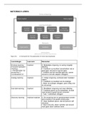
Purification techniques: chromatography (1), electrophoresis (2)
Characterization: mass spectrometry (1), spectroscopy (2), bioassays (3)
1. Chromatography - Separation (polarity, size, charge, boiling point) and purification
2. Electrophoresis - separation (charge-mass ratio)
Charge is based on pI of protein and pH of solution
SDS page: SDS → protein negative charge
3. Mass spectrometry - separation based on mass-to-charge ratio (MW) = most
powerful!
Also differences in post-translational modifications visible
4. Spectroscopy - information about secondary and tertiary structure
Low res.: CD, FTIR, Fluorescence
High res.: NMR, x-ray
diffraction
Bioassays:
- Quantification of small amounts of
proteins
- ELISA (enzyme-linked
immunosorbent assay)
- SPR (surface plasmon resonance)
binding assay
Cell systems
- in vitro biological response
(=mechanism of action)
4
Innovative Therapeutics
1. Structure and stability of proteins (1-9-2025, Trombetta Lima) 2
2. Production of recombinant proteins (2-9-2025, Trombetta Lima) 5
3. Molecular biotechnology and genomics (3-9-2025, Wen Wu) 8
4. Biotherapeutics (4-9-2025, Sousa) 12
5. Development of RNA-based vaccines (5-9-2025, Sousa) 14
6. Molecular basis for the use of light in medicine (8-9-2025, Szymanski) 17
7. Biosimilars and ATMP’s (9-9-2025, Kosterink) 19
8. Immunogeniciteit van therapeutische eiwitten (10-9-2025, Melgert) 21
9. Nanomedicine (11-9-2025, Salvati) 25
10. Innovative therapeutics in de praktijk 1 (11-9-2-2025, Jansman) 28
11. Innovative therapeutics in de praktijk 2 (12-9-2025, Jansman) 30
12. Gene therapy (14-9-2025, Wu) 32
13. Vaccines (15-9-2025, Rafie) 35
, 2
1. Structure and stability of proteins (1-9-2025, Trombetta Lima)
General introduction
Therapeutic proteins are always produced in natural systems (living cells), and are
Macromolecules (1 to >150 kDa) (1st: dyfteria antitoxin = polyclonal ab)
incl.: Biopharmaceuticals, biologicals, biologics,recombinant proteins
Recombinant proteins → use codifying part of DNA and insert in a system that
normally does not produce this protein (1st: insulin in e.coli)
note: Therapeutic peptides are not always biologicals, can be produced synthetically
Therapeutic options: replace a protein that is impaired (1) or block/inactivate a protein (2),
depending on the disease.
Monoclonal antibodies are mainly used in oncology and autoimmune diseases, followed by
infectious diseases.
2
, 3
Structures of proteins
Primary structure: amino acids covalently bound with
peptide bonds → protein backbone
Side chains responsible for properties (MW,
charge (depends on pH/pI), polarity, pka)
Secondary structure
Local folding due to formation hydrogen bonds:
N-H is hydrogen donor
C=O is hydrogen acceptor
a helix: hydrophobic inwards, hydrophilic outside
each turn is 3.4 amino acids long
Tertiary structure: Spatial arrangement of different secondary structures
note: peptides (2-50 amino acids) no tertiary structure
Quaternary structure: Assembly of a stoichiometric fixed number of tertiary molecules into
quaternary one
- mainly non-covalent interactions, can be stabilised by covalent disulfide bridges
Forces of attraction and repulsion:
covalent = peptide bonds, disulfide bridges
non-covalent =
- hydrophobic interactions (nonpolar groups),
- hydrogen bridges (H=electropositive, O/N= electronegative),
- electrostatic interactions (opposite charge),
- van der waals interactions (between dipoles)
Water is bound to proteins → Exterior: from the environment, quite 'loose', Inside ('trapped'):
more strongly bound
Post-translational modifications:
In ER and Golgi-apparatus
(also: formation of disulfide bridges)
3
, 4
Stability of proteins
Folded state: hydrophobic regions on the inside of globular structure, this is
thermodynamically the most favourable (shielded from aqueous environment)
Unfolded/denatured: hydrophobic regions are exposed, thus interactions with environment
which leads to aggregation = clumping of proteins and peptides (and precipitation in
solutions)
Most common chemical reaction during degradation of proteins = oxidation and exchange
(rearrangement) of disulfide bridges, but also hydrolysis of different cysteine units,
deamination and racemization of amino acid residues.
Improve stability of proteins with: genetic engineering (1), replacing reactive amino acid
groups with less reactive ones (=chemical stability) (2), substitute amino acids to improve
intramolecular interactions (=physical stability) (3).
Characterization of proteins
Why? determine: structure, function, purity
When? During development, production and storage
Purification techniques: chromatography (1), electrophoresis (2)
Characterization: mass spectrometry (1), spectroscopy (2), bioassays (3)
1. Chromatography - Separation (polarity, size, charge, boiling point) and purification
2. Electrophoresis - separation (charge-mass ratio)
Charge is based on pI of protein and pH of solution
SDS page: SDS → protein negative charge
3. Mass spectrometry - separation based on mass-to-charge ratio (MW) = most
powerful!
Also differences in post-translational modifications visible
4. Spectroscopy - information about secondary and tertiary structure
Low res.: CD, FTIR, Fluorescence
High res.: NMR, x-ray
diffraction
Bioassays:
- Quantification of small amounts of
proteins
- ELISA (enzyme-linked
immunosorbent assay)
- SPR (surface plasmon resonance)
binding assay
Cell systems
- in vitro biological response
(=mechanism of action)
4