SOFT TISSUE PATHOLOGY
Robbins and Cotran Patho Basis of Disease 10th Ed.
*use at your own risk
TRANS BY: CE
● Soft tissue - refers to non-epithelial tissue excluding the skeleton, joints, Generalizations made based on karyotypic complexity:
central nervous system, and hematopoietic and lymphoid tissues ● Simple karyotype (20%)
● Benign soft tissue tumors are 100-fold more frequent than their malignant ○ Sarcomas are occasionally euploid tumors with a single or limited number of chromosomal changes
counterparts ○ occasionally euploid tumors with a single or limited number of chromosomal changes
● Sarcomas- 2% of all cancer mortality, with aggressive behavior and ○ specific enough to serve as diagnostic markers
resistance to chemotherapy ○ most commonly arise in younger individuals
● Most soft tissue tumors arise in the extremities, particularly the thigh ○ monomorphic microscopic appearance
● 15% arise in children; increases with age ○ E.g Ewing sarcoma and synovial sarcoma
● Most sarcomas are sporadic, no known predisposing cause ● Complex karyotype (80%)
● Sarcoma precursors are undefined ○ usually aneuploid or polyploid
● All highly aggressive malignancies are classified as sarcomas, ○ multiple chromosomal gains and losses
○ producing genomic instability
○ E.g leiomyosarcomas and undifferentiated pleomorphic sarcoma
○ more common in adults
○ microscopically diverse (pleomorphic)
CATEGORY TUMOR TYPE BEHAVIOR COMMON AGE MORPHOLOGY CLINICAL FEATURES PROGNOSIS
LOCATION
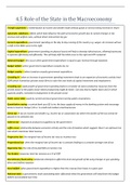
ADIPOSE LIPOMA Benign Superficial 40–60 - Mature adipose tissue - most common soft tissue tumor in adults cured by simple excision
proximal middle - well-encapsulated - soft, mobile, painless,
extremity, trunk adulthood - arises in the subcutis
- large, intramuscular, and poorly circumscribed
- Lipomatosis - multifocal lipomas involve a limb
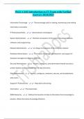
LIPOSARCOMA
- most common sarcomas of adulthood
- malignant
- typically develop in deep soft tissues of proximal extremities and retroperitoneum
- sixth and seventh decades
- recur locally, and often repeatedly, unless adequately excised
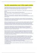
WELL-DIFFERENTIATED Deep extremity, 50–60 - Adipose tissue with scattered atypical spindle cells - amplifications of chromosome region indolent
LIPOSARCOMA retroperitoneum 12q13–q15, includes p53 inhibitor MDM2
-
MYXOID LIPOSARCOMA Thigh, leg 30s - Myxoid matrix, “chicken wire” vessels, round cells, - fusion gene generated by a t(12;16) intermediate
lipoblasts translocation
Malignant
- abundant basophilic extracellular matrix, arborizing
capillaries, and primitive cells at various stages of
adipocyte differentiation resembling fetal fat
PLEOMORPHIC - sheets of anaplastic cells with bizarre nuclei admixed - complex karyotypes without reproducible aggressive and frequently
LIPOSARCOMA - variable numbers of immature adipocytes - lipoblast genetic abnormalities metastasize
1
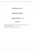
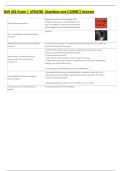
, FIBROUS NODULAR FASCIITIS Arm, forearm, 20–30 - Tissue culture growth, extravasated erythrocytes - self-limited fibroblastic and myofibroblastic regresses spontaneously
chest and back young - arises in the deep dermis, subcutis, fascia, or muscle proliferation if excised, rarely recurs
adults - non-encapsulated - grow rapidly over weeks to months
- well-circumscribed or slightly infiltrative - history of trauma in 10% to 50% of case -
- less than 3 cm diameter previously considered a reactive lesion
- plump, immature-appearing fibroblasts and - clonal proliferation t(17;22) translocation,
myofibroblasts containing elongated nuclei with punctate produces MYH9-USP6 fusion
nucleoli - defect that prevents neoplastic cells to be
- Mitoses are frequent, but atypical forms are notably malignant - not defined
absent
- Zonation - gradient, hypercellular regions with myxoid
stroma to hypocellular areas with fibrous stroma
- Storiform or fascicular patterns - common in cellular areas
- Metaplastic bone, cystic areas, ganglion like cells,
prominent vessels, extravasated red cells, and infiltrating
lymphocytes are common
SUPERFICIAL - plump spindle cells arranged in poorly defined broad - can cause local deformity but has an - Palmar and plantar
FIBROMATOSIS bundles or long, sweeping fascicles surrounded by innocuous clinical course fibromatoses progress in
abundant dense collagen - affects males more about 50% of cases
- remainder stabilize and do
SUBTYPES: not progress
● Dupuytren contracture or palmar - some resolve
fibromatosis spontaneously
○ irregular or nodular thickening of the - recurrence is common
Benign palmar fascia even after excision
○ unilateral or bilateral
○ Incidence increases with age
○ puckering or dimpling and a slowly
progressive flexion contracture’
● Ledderhose disease or plantar fibromatosis
○ boys from under 10 years of age into
adolescence
○ Unilateral
○ does not cause contractures
○ can be associated with palmar and penile
fibromatosis
● Peyronie disease or penile fibromatosis
○ palpable induration or mass on the
dorsolateral aspect of the penis
○ may cause abnormal curvature of the
shaft and constriction of the urethra
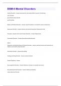
DEEP FIBROMATOSIS musculoaponeur 30–40 - Bland fibroblast, Dense collagen, long, parallel - also called Desmoid tumors - frequently recur but do
otic structures of unidirectional fascicles - large, infiltrative masses not metastasize
the anterior - gray-white, firm, poorly demarcated masses - predominantly in women - possibly disfiguring or
Abdominal wall - 1 to 15 cm diameter - can also arise in the limb girdles or mesentery disabling
- rubbery and tough - mutations in the APC or CTNNB1 (β-catenin) - complete excision can be
- infiltrate surrounding muscle, nerve, and fat genes, leading to increased Wnt signaling difficult
- histologic appearance can resemble a scar - majority - sporadic CTNNB1 mutations
2
Robbins and Cotran Patho Basis of Disease 10th Ed.
*use at your own risk
TRANS BY: CE
● Soft tissue - refers to non-epithelial tissue excluding the skeleton, joints, Generalizations made based on karyotypic complexity:
central nervous system, and hematopoietic and lymphoid tissues ● Simple karyotype (20%)
● Benign soft tissue tumors are 100-fold more frequent than their malignant ○ Sarcomas are occasionally euploid tumors with a single or limited number of chromosomal changes
counterparts ○ occasionally euploid tumors with a single or limited number of chromosomal changes
● Sarcomas- 2% of all cancer mortality, with aggressive behavior and ○ specific enough to serve as diagnostic markers
resistance to chemotherapy ○ most commonly arise in younger individuals
● Most soft tissue tumors arise in the extremities, particularly the thigh ○ monomorphic microscopic appearance
● 15% arise in children; increases with age ○ E.g Ewing sarcoma and synovial sarcoma
● Most sarcomas are sporadic, no known predisposing cause ● Complex karyotype (80%)
● Sarcoma precursors are undefined ○ usually aneuploid or polyploid
● All highly aggressive malignancies are classified as sarcomas, ○ multiple chromosomal gains and losses
○ producing genomic instability
○ E.g leiomyosarcomas and undifferentiated pleomorphic sarcoma
○ more common in adults
○ microscopically diverse (pleomorphic)
CATEGORY TUMOR TYPE BEHAVIOR COMMON AGE MORPHOLOGY CLINICAL FEATURES PROGNOSIS
LOCATION
ADIPOSE LIPOMA Benign Superficial 40–60 - Mature adipose tissue - most common soft tissue tumor in adults cured by simple excision
proximal middle - well-encapsulated - soft, mobile, painless,
extremity, trunk adulthood - arises in the subcutis
- large, intramuscular, and poorly circumscribed
- Lipomatosis - multifocal lipomas involve a limb
LIPOSARCOMA
- most common sarcomas of adulthood
- malignant
- typically develop in deep soft tissues of proximal extremities and retroperitoneum
- sixth and seventh decades
- recur locally, and often repeatedly, unless adequately excised
WELL-DIFFERENTIATED Deep extremity, 50–60 - Adipose tissue with scattered atypical spindle cells - amplifications of chromosome region indolent
LIPOSARCOMA retroperitoneum 12q13–q15, includes p53 inhibitor MDM2
-
MYXOID LIPOSARCOMA Thigh, leg 30s - Myxoid matrix, “chicken wire” vessels, round cells, - fusion gene generated by a t(12;16) intermediate
lipoblasts translocation
Malignant
- abundant basophilic extracellular matrix, arborizing
capillaries, and primitive cells at various stages of
adipocyte differentiation resembling fetal fat
PLEOMORPHIC - sheets of anaplastic cells with bizarre nuclei admixed - complex karyotypes without reproducible aggressive and frequently
LIPOSARCOMA - variable numbers of immature adipocytes - lipoblast genetic abnormalities metastasize
1
, FIBROUS NODULAR FASCIITIS Arm, forearm, 20–30 - Tissue culture growth, extravasated erythrocytes - self-limited fibroblastic and myofibroblastic regresses spontaneously
chest and back young - arises in the deep dermis, subcutis, fascia, or muscle proliferation if excised, rarely recurs
adults - non-encapsulated - grow rapidly over weeks to months
- well-circumscribed or slightly infiltrative - history of trauma in 10% to 50% of case -
- less than 3 cm diameter previously considered a reactive lesion
- plump, immature-appearing fibroblasts and - clonal proliferation t(17;22) translocation,
myofibroblasts containing elongated nuclei with punctate produces MYH9-USP6 fusion
nucleoli - defect that prevents neoplastic cells to be
- Mitoses are frequent, but atypical forms are notably malignant - not defined
absent
- Zonation - gradient, hypercellular regions with myxoid
stroma to hypocellular areas with fibrous stroma
- Storiform or fascicular patterns - common in cellular areas
- Metaplastic bone, cystic areas, ganglion like cells,
prominent vessels, extravasated red cells, and infiltrating
lymphocytes are common
SUPERFICIAL - plump spindle cells arranged in poorly defined broad - can cause local deformity but has an - Palmar and plantar
FIBROMATOSIS bundles or long, sweeping fascicles surrounded by innocuous clinical course fibromatoses progress in
abundant dense collagen - affects males more about 50% of cases
- remainder stabilize and do
SUBTYPES: not progress
● Dupuytren contracture or palmar - some resolve
fibromatosis spontaneously
○ irregular or nodular thickening of the - recurrence is common
Benign palmar fascia even after excision
○ unilateral or bilateral
○ Incidence increases with age
○ puckering or dimpling and a slowly
progressive flexion contracture’
● Ledderhose disease or plantar fibromatosis
○ boys from under 10 years of age into
adolescence
○ Unilateral
○ does not cause contractures
○ can be associated with palmar and penile
fibromatosis
● Peyronie disease or penile fibromatosis
○ palpable induration or mass on the
dorsolateral aspect of the penis
○ may cause abnormal curvature of the
shaft and constriction of the urethra
DEEP FIBROMATOSIS musculoaponeur 30–40 - Bland fibroblast, Dense collagen, long, parallel - also called Desmoid tumors - frequently recur but do
otic structures of unidirectional fascicles - large, infiltrative masses not metastasize
the anterior - gray-white, firm, poorly demarcated masses - predominantly in women - possibly disfiguring or
Abdominal wall - 1 to 15 cm diameter - can also arise in the limb girdles or mesentery disabling
- rubbery and tough - mutations in the APC or CTNNB1 (β-catenin) - complete excision can be
- infiltrate surrounding muscle, nerve, and fat genes, leading to increased Wnt signaling difficult
- histologic appearance can resemble a scar - majority - sporadic CTNNB1 mutations
2