CASE 1
1. NEURAL DEVELOPMENT
STAGES OF PRENATAL DEVELOPMENT
1. Germinal stage (weeks 1-2)
o Fertilization and zygote formation
o Implantation → placenta and amniotic sac
2. Embryonic stage (week 3-8)
o Gastrulation (week 3-4)
o Neurulation (week 4-5)
o Organogenesis (week 6)
3. Fetal stage (week 9 till birth)
o Brain and nervous system maturation
o Bone development and growth
o Organ functionality
GASTRULATION
PROCESS OF GASTRULATION
Day 14-15
1. Primitive groove formation → narrow
depression into center of epiblast layer
o Hypoblast → yolk sac
o Epiblast → amniotic cavity
o Prechordal plate → area
columnar cells
o The cells of the epiblast (on top of
the hypoblast) migrate inwards,
downwards and then
differentiate. → primitive streak
- Defines all major body axis and
is composed of node, pit and
groove.
- Cranial-caudal
- Dorsal-ventral
, 2. Primitive node and pit → at the cranial end of the streak a thick cluster of cells creates the
primitive node → depression called primitive pit → surrounded with slightly elevated ectoderm.
3. Layer formation
o Endoderm: formed as the migrating cells replace the hypoblast
o → tissues and organs
o Ectoderm: derived from remaining epiblast cells, differentiates into structures like the
neural plate.
→ nervous system
o Mesoderm: emerge between the ectoderm and endoderm as cells force their way
inward, completeing the trilaminar structure.
- → paraxial (central), Intermediate (gonads and kidneys), lateral plate mesoderm
(splanchic and somatic).
Day 17: notochord formation
o A rod-like structure called the notochord → forms from mesenchymal cells.
o This temporary structure aids in shaping the embryo’s body by
guiding it’s folding.
→ SHH secretion → tissue differentiation.
Day 20: mesoderm differentiation and neurulation
o Mesoderm cells surrounding the notochord differentiate into
three distinct layers
1. paraxial mesoderm: vertebrae and skeletal muscle
2. intermediate mesoderm: urogenital system
3. lateral plate mesoderm: circulatory system, body wall, limb
structures.
→ notochord neurulation → ectoderm to form the neural plate →
nervous system.
End of week 3 → formation of bilaminar membranes
o Two specialized bilaminar regions form within the trilaminar disc
o Cranial bilaminar region (oropharyngeal membrane) → mouth
o Caudal bilaminar region (cloacal membrane) → anus and genitourinary structures
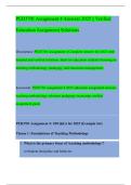
,NEURULATION
Day 20: formation of the neural groove and neural tube
1. Neural plate formation
2. Neural groove formation: neural plate → neural
groove → neural folds on the side
3. Fusion of neural folds → fuse → open ends →
cranial neuropore and caudal neuropore (will
eventually close)
4. Formation of notochord and neural tube
o Neural crest cells → resides ate the peaks of
neural folds → folds fuse → layer over the closed
neural tube → differentiate → peripheral nervous
system → neurons, glia and parts of the autonomic
nervous system.
FIGURE 1: NEURULATION
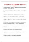
, FORMATION OF THE BRAIN
o Formation of the brain begins with the development of three primary vesicles → three-
vesicle stage → shape to brain structure → differentiate into five distinct brain regions.
1. Prosencephalon (forebrain): the most anterior (front) part of the developing brain
2. Mesencephalon (midbrain): located between the forebrain and the hindbrain, responsible for
sensory processing and movement.
3. Rhombencephalon (hindbrain): The posterior part, giving rise to structures that control basic
functions like heart rate and balance.
Five-vesicle stage → vesicles further divide
o Prosencephalon → telencephalon and diencephalon
o Mesencephalon → single vescicle
o Rhombencephalon → metencephalon and myelencephalon
• Proliferate and differentiate
o Telencephalon → cortex
o Neurons cognition, sensory perception, and motor control.
The brain develops from the walls of the five fluid-filled vesicles, in the early stages consisting of two
layers:
o Ventricular zone
o Marginal zone
1. NEURAL DEVELOPMENT
STAGES OF PRENATAL DEVELOPMENT
1. Germinal stage (weeks 1-2)
o Fertilization and zygote formation
o Implantation → placenta and amniotic sac
2. Embryonic stage (week 3-8)
o Gastrulation (week 3-4)
o Neurulation (week 4-5)
o Organogenesis (week 6)
3. Fetal stage (week 9 till birth)
o Brain and nervous system maturation
o Bone development and growth
o Organ functionality
GASTRULATION
PROCESS OF GASTRULATION
Day 14-15
1. Primitive groove formation → narrow
depression into center of epiblast layer
o Hypoblast → yolk sac
o Epiblast → amniotic cavity
o Prechordal plate → area
columnar cells
o The cells of the epiblast (on top of
the hypoblast) migrate inwards,
downwards and then
differentiate. → primitive streak
- Defines all major body axis and
is composed of node, pit and
groove.
- Cranial-caudal
- Dorsal-ventral
, 2. Primitive node and pit → at the cranial end of the streak a thick cluster of cells creates the
primitive node → depression called primitive pit → surrounded with slightly elevated ectoderm.
3. Layer formation
o Endoderm: formed as the migrating cells replace the hypoblast
o → tissues and organs
o Ectoderm: derived from remaining epiblast cells, differentiates into structures like the
neural plate.
→ nervous system
o Mesoderm: emerge between the ectoderm and endoderm as cells force their way
inward, completeing the trilaminar structure.
- → paraxial (central), Intermediate (gonads and kidneys), lateral plate mesoderm
(splanchic and somatic).
Day 17: notochord formation
o A rod-like structure called the notochord → forms from mesenchymal cells.
o This temporary structure aids in shaping the embryo’s body by
guiding it’s folding.
→ SHH secretion → tissue differentiation.
Day 20: mesoderm differentiation and neurulation
o Mesoderm cells surrounding the notochord differentiate into
three distinct layers
1. paraxial mesoderm: vertebrae and skeletal muscle
2. intermediate mesoderm: urogenital system
3. lateral plate mesoderm: circulatory system, body wall, limb
structures.
→ notochord neurulation → ectoderm to form the neural plate →
nervous system.
End of week 3 → formation of bilaminar membranes
o Two specialized bilaminar regions form within the trilaminar disc
o Cranial bilaminar region (oropharyngeal membrane) → mouth
o Caudal bilaminar region (cloacal membrane) → anus and genitourinary structures
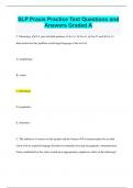
,NEURULATION
Day 20: formation of the neural groove and neural tube
1. Neural plate formation
2. Neural groove formation: neural plate → neural
groove → neural folds on the side
3. Fusion of neural folds → fuse → open ends →
cranial neuropore and caudal neuropore (will
eventually close)
4. Formation of notochord and neural tube
o Neural crest cells → resides ate the peaks of
neural folds → folds fuse → layer over the closed
neural tube → differentiate → peripheral nervous
system → neurons, glia and parts of the autonomic
nervous system.
FIGURE 1: NEURULATION
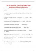
, FORMATION OF THE BRAIN
o Formation of the brain begins with the development of three primary vesicles → three-
vesicle stage → shape to brain structure → differentiate into five distinct brain regions.
1. Prosencephalon (forebrain): the most anterior (front) part of the developing brain
2. Mesencephalon (midbrain): located between the forebrain and the hindbrain, responsible for
sensory processing and movement.
3. Rhombencephalon (hindbrain): The posterior part, giving rise to structures that control basic
functions like heart rate and balance.
Five-vesicle stage → vesicles further divide
o Prosencephalon → telencephalon and diencephalon
o Mesencephalon → single vescicle
o Rhombencephalon → metencephalon and myelencephalon
• Proliferate and differentiate
o Telencephalon → cortex
o Neurons cognition, sensory perception, and motor control.
The brain develops from the walls of the five fluid-filled vesicles, in the early stages consisting of two
layers:
o Ventricular zone
o Marginal zone