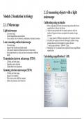
.1.3 measuring objects with a light
2
Module 2 foundation in biology microscope
Calibrating using graticules
2.1.1 Microscope 1. P lace a stage graticule on the microscope stage and use the lowest
magnification to observe the division
Light microscope 2. Align the stage graticule and the eyepiece graticule, check the
- 2 D colour image number of eyepiece division correspond to the number of stage
- Limited magnification and resolution graticule
- Can see nuclei, but not ribosomes, endoplasmic reticulum lysosomes -> 1 stage graticule (1000µm) corresponds to 40 eyepiece divisions
3. Calculate each eyepiece division by dividing the number of stage
Laser scanning confocal microscope graticule division over the number of eyepiece division
- 3 D colour image -> each eyepiece division = 1000/40 = 25µm
- Image is high resolution and high contrast 4. Repeat step 1 to 3 to calculate each eyepiece division using different
- Cells must be stained with fluorescent dyes for observation magnification
- It has depth selectivity, which can focus on structures at different
depths
Calculating magnification (I AM)
Transmission electron microscope (TEM)
- 2 D black and white image
- Organelles can be observed
- Cannot observe live specimens
Scanning electron microscope (SEM)
- 3 D black and white image
- Image is low resolution
- False colour can be added by computer
- Surface of specimens can be observed
,