1
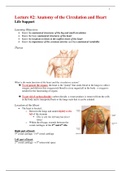
Lecture #2: Anatomy of the Circulation and Heart
Life Support
Learning Objectives
• Know the anatomical structures of the big and small circulation
• Know the basic anatomical structures of the heart
• Know the locations to listen to the outflow tracts of the heart
• Know the importance of the coronary arteries and their anatomical variability
Thorax
What is the main function of the heart and the circulatory system?
➔ To oxygenate the organs: the heart is the “pump” that sends blood to the lungs to collect
oxygen, and delivers this oxygen-rich blood to every organ/cell in the body → oxygen is
needed for the functioning of organs
➔ To get rid of carbon dioxide: carbon dioxide, a waste product, is removed from the cells
in the body and is transported back to the lungs such that it can be exhaled
Location of the Heart
• The heart is located…
o Between the lungs and under/slightly to the
left of the sternum
▪ This is why the left lung has fewer
lobes!
o Within the ribcage; extends between the
costal cartilages of the 2nd and 6th ribs
Right part of heart:
3rd costal cartilage → 6th costal cartilage
Left part of heart:
2nd costal cartilage → 5th intercostal space
, 2
Mediastinum
• Central, middle compartment of the thoracic cavity,
enclosed on the right and left by the pleurae
The mediastinum is composed of:
1) Thymus:
➔ Only children have this structure; disappears in
adults
o “After puberty, the thymus starts to slowly
shrink and becomes replaced by fat”
➔ A small, irregularly-shaped organ that is part of the
lymphatic system
➔ Produces T cells (T lymphocytes), which defend
against pathogens (e.g. bacteria, viruses), and are important to the immune system
2) Pericard:
➔ A double-layered sac of tissue that surrounds the heart and the roots of the great
blood vessels
Mediastinum
• The sternal plane (angulus ludovici) divides the mediastinum in
superior and inferior compartments
• The inferior compartment is divided into 3 sections:
o Inferior anterior mediastinum:
▪ The space between the sternum and the pericard/heart
o Inferior middle mediastinum:
▪ Where the heart is located
o Inferior posterior mediastinum:
▪ The space behind the heart, to the vertebrae
▪ Contains the aorta, esophagus, etc.
, 3
Mediastinum Inferior
• The mediastinum inferior is divided into three compartments:
1. Anterior
2. Medium
3. Posterior
Mediastinum Medium
• The inferior middle mediastinum contains:
o The pericard, which covers the:
▪ Heart
▪ Roots of the great vessels
• The outflow tracts of the great vessels are in
the pericard
• CAVE
o Outside pericard is the N. Phrenicus (2x)
▪ The nerve that innervates the diaphragm
▪ Sends signals to the diaphragm to either contract (i.e.
move down) during inhalation or relax (i.e. move up)
during exhalation
The right phrenic nerve descends on the right side of the right atrium
(separated from it by the pericard)
The left phrenic nerve descends on the left side of the left ventricle (separated
from it by the pericard)
Pericard
• Can be divided into:
o Fibrous pericardium
▪ Most superficial/outer layer of the
pericardium; very stiff
▪ Made of connective tissue, and provides
support and protection for the heart
o Serous pericardium (divided into two layers)
▪ Parietal pericardium
• Fused into and inseparable from
the fibrous pericardium
▪ Visceral pericardium
• The inner layer, which outlines
the outer surface of the heart itself
• Between the two layers of the serous pericardium is the pericardial cavity, which
contains pericardial fluid
o This fluid provides lubrication between the layers, and allows the heart to expand
and contract