Case 1 – the developing nervous system
Learning Goals:
1. Recap prenatal development of the nervous system.
a. Neurulation, cell migration, cell types
2. How do stem cells differentiate?
a. Notch signaling
3. How do synapses form, work, and how are they degenerated?
a. Use figure 3, neurotrophic signaling, neurotrophic factor hypothesis, synaptic
plasticity
b. Difference between CNS and PNS
4. What are the stages of postnatal development?
a. Use figure 4
5. What happens to nerve cells after damage (e.g. physical trauma not disease related)?
a. Regeneration, generation of new cells?
b. Difference between CNS and PNS
, 1. Recap prenatal development of the nervous system.
a. Neurulation, cell migration, cell types
After gastrulation, when the three-laminar embryo is formed (endoderm, mesoderm, ectoderm), of
which the ectoderm will differentiate into the nervous system (and skin). After gastrulation, the
notochord is formed from the mesoderm which sends signals to the overlying ectoderm, inducing it
to become neuroectoderm. This results in a strip of neuronal stem cells that runs along the back of
the foetus called the neural plate, it is the origin of the entire nervous system.
The neural plate folds outwards to form the neural groove, beginning in the neck region the neural
folds of the groove close to create the neural tube. This form of neurulation is called the primary
neurulation.
The anterior part of the neural tube is called the basal plate and the posterior part is called the alar
plate. The hollow interior is called the neural canal. At the end of the fourth week, the open ends of
the neural tube close off.
The neural tube gets divided in the forebrain (prosencephalon), midbrain (mesencephalon), hindbrain
(rhombencephalon) and the spinal cord. After the brain divisions are marked by expansions of the
neural tube, they are marked by expansions of the neural tube called primary brain vesicles.
During the 5th week, the mesencephalon becomes larger and the prosencephalon and
rhombencephalon divide in 2 portions:
, • Prosencephalon -> telencephalon and diencephalon
• Mesencephalon -> mesencephalon
• Rhombencephalon -> metencephalon and myelencephalon
The PNS if formed from the neural crest cells which is neural ectoderm lateral of the neural tube.
Ventricles -> CSF
In ventricular zone -> cells duplicate
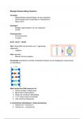
, 2. How do stem cells differentiate?
a. Notch signaling
Neural structures develop in three major steps:
1. Proliferation
2. Migration
3. Differentiations
five positions??
Cell proliferation is the addition of new stem cells through mitosis which happens as soon as the
neural tube closes. The neural stem cells are located in the ventricular zone and their proliferation
can be either symmetrical or asymmetrical:
• Symmetrical cell division: both daughter cells remain in the ventricular zone and undergo
further proliferation. The two new stem cells are identical. This is called vertical cleavage
because the cells stay in the same plane as the ventricular zone.
• Asymmetric cell division: one daughter cell stays in the ventricular zone while the other one
migrates towards the pial surface. This gives rise to non-identical daughter cells. One
becomes a neural stem cell and the other becomes a neural precursor and starts to migrate
and differentiate. This is called horizontal cleavage.
Important picture for exam
Learning Goals:
1. Recap prenatal development of the nervous system.
a. Neurulation, cell migration, cell types
2. How do stem cells differentiate?
a. Notch signaling
3. How do synapses form, work, and how are they degenerated?
a. Use figure 3, neurotrophic signaling, neurotrophic factor hypothesis, synaptic
plasticity
b. Difference between CNS and PNS
4. What are the stages of postnatal development?
a. Use figure 4
5. What happens to nerve cells after damage (e.g. physical trauma not disease related)?
a. Regeneration, generation of new cells?
b. Difference between CNS and PNS
, 1. Recap prenatal development of the nervous system.
a. Neurulation, cell migration, cell types
After gastrulation, when the three-laminar embryo is formed (endoderm, mesoderm, ectoderm), of
which the ectoderm will differentiate into the nervous system (and skin). After gastrulation, the
notochord is formed from the mesoderm which sends signals to the overlying ectoderm, inducing it
to become neuroectoderm. This results in a strip of neuronal stem cells that runs along the back of
the foetus called the neural plate, it is the origin of the entire nervous system.
The neural plate folds outwards to form the neural groove, beginning in the neck region the neural
folds of the groove close to create the neural tube. This form of neurulation is called the primary
neurulation.
The anterior part of the neural tube is called the basal plate and the posterior part is called the alar
plate. The hollow interior is called the neural canal. At the end of the fourth week, the open ends of
the neural tube close off.
The neural tube gets divided in the forebrain (prosencephalon), midbrain (mesencephalon), hindbrain
(rhombencephalon) and the spinal cord. After the brain divisions are marked by expansions of the
neural tube, they are marked by expansions of the neural tube called primary brain vesicles.
During the 5th week, the mesencephalon becomes larger and the prosencephalon and
rhombencephalon divide in 2 portions:
, • Prosencephalon -> telencephalon and diencephalon
• Mesencephalon -> mesencephalon
• Rhombencephalon -> metencephalon and myelencephalon
The PNS if formed from the neural crest cells which is neural ectoderm lateral of the neural tube.
Ventricles -> CSF
In ventricular zone -> cells duplicate
, 2. How do stem cells differentiate?
a. Notch signaling
Neural structures develop in three major steps:
1. Proliferation
2. Migration
3. Differentiations
five positions??
Cell proliferation is the addition of new stem cells through mitosis which happens as soon as the
neural tube closes. The neural stem cells are located in the ventricular zone and their proliferation
can be either symmetrical or asymmetrical:
• Symmetrical cell division: both daughter cells remain in the ventricular zone and undergo
further proliferation. The two new stem cells are identical. This is called vertical cleavage
because the cells stay in the same plane as the ventricular zone.
• Asymmetric cell division: one daughter cell stays in the ventricular zone while the other one
migrates towards the pial surface. This gives rise to non-identical daughter cells. One
becomes a neural stem cell and the other becomes a neural precursor and starts to migrate
and differentiate. This is called horizontal cleavage.
Important picture for exam