CET Exam Questions With 100%
Verified Correct Answers.
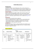
ST segment elevation may indicate
Myocardial Infarction
Difficulty breathing
dyspnea
A pacing spike followed by a P wave shows a:
Atrial pacemaker
To more easily view a tachycardia rhythm, you can:
Increase the machine speed
4th ICS, Right Sternal border
V1
The vertical axis of the ECG graph paper represents:
Voltage (gain)
"Pacemaker" of the heart; Electrical impulse begins here
SA node
T wave inversion may indicate
Ischemia
Calculate a target heart rate for an exercise stress test by using the following formula:
220 minus the patient's age
, Reasons to terminate cardiac stress testing
Chest pain;
moderate to severe SOB;
dizziness;
B/P changes;
severe fatigue;
cold, clammy skin
Shows from the beginning of atrial depolarization to the beginning of
ventricular depolarization
PR interval
4th ICS, Left sternal border
V2
Artifact due to patient movement such as tremors:
Somatic tremor
Methods for calculation of the heart rate
300 divided by the number of large squares between R waves (Sequence
300/150/100/75/60/50); 1500 divided bu the number of small squares between R waves;
Number of R waves in a 6-second strip times 10.
Standard ECG sensitivity
10mm/1mV
(2 large boxes)
Records RA - LA
Lead I
Temporary lack of blood flow to the heart
ischemia
Verified Correct Answers.
ST segment elevation may indicate
Myocardial Infarction
Difficulty breathing
dyspnea
A pacing spike followed by a P wave shows a:
Atrial pacemaker
To more easily view a tachycardia rhythm, you can:
Increase the machine speed
4th ICS, Right Sternal border
V1
The vertical axis of the ECG graph paper represents:
Voltage (gain)
"Pacemaker" of the heart; Electrical impulse begins here
SA node
T wave inversion may indicate
Ischemia
Calculate a target heart rate for an exercise stress test by using the following formula:
220 minus the patient's age
, Reasons to terminate cardiac stress testing
Chest pain;
moderate to severe SOB;
dizziness;
B/P changes;
severe fatigue;
cold, clammy skin
Shows from the beginning of atrial depolarization to the beginning of
ventricular depolarization
PR interval
4th ICS, Left sternal border
V2
Artifact due to patient movement such as tremors:
Somatic tremor
Methods for calculation of the heart rate
300 divided by the number of large squares between R waves (Sequence
300/150/100/75/60/50); 1500 divided bu the number of small squares between R waves;
Number of R waves in a 6-second strip times 10.
Standard ECG sensitivity
10mm/1mV
(2 large boxes)
Records RA - LA
Lead I
Temporary lack of blood flow to the heart
ischemia