Case 1. The organization of the
brain
The brain is the most complex organ of the human body and, although it has been extensively studied, the exact
workings remain elusive. In the early days of neuroscience, when there was no modern technology available, individual
cases of brain injury (e.g. by trauma) gave valuable insight into the workings of the brain. Some historical examples:
1. The Edwin Smith Papyrus is an ancient Egyptian medical text, named after the dealer who bought it in 1862,
and the oldest known surgical treatise on trauma. This document, which may have been a manual of military
surgery, describes 48 cases and gives the first accounts of several forms of brain injury and their associated
complications. A quote from Case 20 from the Edwin Smith Papyrus scrolls: ‘’thou examinest a man having a
wound in his temple, penetrating to the bone, (and) perforating his temporal bone, if thou ask of him
concerning his malady and he speak not to thee’’
2. Ernest Aubertin, a young scientist in the 19th century came across an interesting case in 1861. The patient
had blown away a big part of his left frontal cranium during a suicide attempt after shooting himself in the
head. While the injury had apparently left both speech and intellect intact, Aubertin discovered he could
abruptly stop the patient in mid-sentence by applying pressure to the exposed frontal lobes with a spatula.
However, speech returned immediately after the compression ceased.
3. Phineas Gage was a young railroad construction foreman in the 19th century. In September 1848, in an
unfortunate accident, an explosion blasted a steel rod through his skull. Most of the damage was done to the
ventromedial region of the frontal lobes of his brain on both sides. He recovered, but months later Gage began
to have startling changes in personality and mood. He became extravagant and anti-social, foul-mouthed and
a liar with bad manners, and could no longer hold a job or plan his future. "Gage was no longer Gage", said his
friends of him.
4. When the German army was battling the advancing Russian Red Army in October 1944, a 36-year-old German
lieutenant on the East Prussian front was hit by shrapnel from an artillery grenade. He was evacuated to a
field hospital, where a surgeon removed a piece of metal from the back of his head. The external wound
healed, but it soon became clear that something strange was going on. The officer reported that he could no
longer see faces.
Occipital (back of head) vision was damaged
These individual cases initiated the development of knowledge of the macroscopic and functional organization of the
brain, including the different subdivisions of the nervous system and the distinct brain regions. But, of course, when we
focus closely on the brain, it is evident that the brain comprises various cell types. These cell types include neurons and
supporting glia cells. Nowadays, we have a wealth of knowledge, including detailed insights into what the different
nervous systems and brain regions do, the structure of neurons and the recognition that synapses exist between them.
In addition, supporting glia cells have different key functions. There are so many details, but let’s begin by getting a
global overview.
Key words:
Parietal lobe, frontal lobes (higher intelligence, language, memory, personality
control), occipital lobe (vision), temporal lobe (temporal bone covering lobe)
Cerebellum
Gyrus (have different names), sulcus need this because it reduces the volume
of the brain
Crania: bone that is covering the brain
Vertebrae: covering spinal cord
Head injury: injury to crania and brain
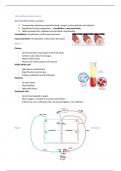
Membranes that cover the brain:
Skin aponeurosis periosteum bone dura materarachnoidBetween this
space, filled with spinal fluid, produced and circulated to the body, pia mater
Meninges (dura mater, arachnoid, pia mater)
Lateral ventricles: where cerebral spinal fluid is produced and circulates
CSF is not only inside but also surrounding
CSF circulation
,Brain: grey (outside) and white matter (inside, fiber) different in spinal cord
(grey matter: inside cell bodies, white matter: outside, extensions)
Latin words!!!
Connective tissue in nervous system: glial cells
Nervous system
Nervous system has 2 parts:
1. Central nervous system
2. Peripheral nervous system
The central nervous system (CNS) is made up of the:
- Brain (cerebrum and cerebellum)
- Spinal cord
The peripheral nervous system is made up of the nerves that branch
off from the spinal cord to the rest of the body
Microscopic: What is the structure of the neuron?
(+ glial cells)
Nervous tissue is composed of 2 types of cells:
1. Neurons= cells that can receive and transmit chemical or
electrical signal
2. Glial cells= cells that provide support functions for the
neurons by playing an information processing role that is
complementary to neurons
Structure neuron:
Cell body (soma): contains nucleus, smooth and rough endoplasmic
reticulum, Golgi apparatus, mitochondria and other cellular components
Dendrites: The receiving part of the neuron. Dendrites receive synaptic
inputs from axons some dendrites have small protrusions=dendritic
spines, which increase surface area for possible synaptic connections
Axons: Long, thin structure in which action potentials are generated, the
transmitting part of the neuron. arises from the cell body at a specialized
area called the axon hillock. Many axons are covered with myelin (helps
them convey the nerve impulse rapidly). Towards the ends of a axon, it
, splits into many branches and develops as axon terminals (make
connections on target cells)
Nerves: bundle of axons
Glial cells functions:
- Responsibility of maintaining a homeostatic balance
- Myelinating neurons
- Providing structural support for neurons
- Protecting neurons
Astrocytes:
- most abundant cells in the brain
- end feet of the astrocytes form the glia limitans on the visceral surface
of the pia mater, by joining multiple layers of end feet this limits the
communications between the CNS and other tissue
- provide physical and nutritional support for neurons (clean up brain
‘debris’, transport nutrients to neurons etc.
Blood-brain barrier
further subdivided into:
- Protoplasmic astrocytes
Located in the grey matter
- Fibrous astrocytes
More commonly found in and are oriented
longitudinally within the plane of the fiber bundles
of the white matter
Oligodendrocytes:
- myelin-forming cells of the CNS, have long
brain
The brain is the most complex organ of the human body and, although it has been extensively studied, the exact
workings remain elusive. In the early days of neuroscience, when there was no modern technology available, individual
cases of brain injury (e.g. by trauma) gave valuable insight into the workings of the brain. Some historical examples:
1. The Edwin Smith Papyrus is an ancient Egyptian medical text, named after the dealer who bought it in 1862,
and the oldest known surgical treatise on trauma. This document, which may have been a manual of military
surgery, describes 48 cases and gives the first accounts of several forms of brain injury and their associated
complications. A quote from Case 20 from the Edwin Smith Papyrus scrolls: ‘’thou examinest a man having a
wound in his temple, penetrating to the bone, (and) perforating his temporal bone, if thou ask of him
concerning his malady and he speak not to thee’’
2. Ernest Aubertin, a young scientist in the 19th century came across an interesting case in 1861. The patient
had blown away a big part of his left frontal cranium during a suicide attempt after shooting himself in the
head. While the injury had apparently left both speech and intellect intact, Aubertin discovered he could
abruptly stop the patient in mid-sentence by applying pressure to the exposed frontal lobes with a spatula.
However, speech returned immediately after the compression ceased.
3. Phineas Gage was a young railroad construction foreman in the 19th century. In September 1848, in an
unfortunate accident, an explosion blasted a steel rod through his skull. Most of the damage was done to the
ventromedial region of the frontal lobes of his brain on both sides. He recovered, but months later Gage began
to have startling changes in personality and mood. He became extravagant and anti-social, foul-mouthed and
a liar with bad manners, and could no longer hold a job or plan his future. "Gage was no longer Gage", said his
friends of him.
4. When the German army was battling the advancing Russian Red Army in October 1944, a 36-year-old German
lieutenant on the East Prussian front was hit by shrapnel from an artillery grenade. He was evacuated to a
field hospital, where a surgeon removed a piece of metal from the back of his head. The external wound
healed, but it soon became clear that something strange was going on. The officer reported that he could no
longer see faces.
Occipital (back of head) vision was damaged
These individual cases initiated the development of knowledge of the macroscopic and functional organization of the
brain, including the different subdivisions of the nervous system and the distinct brain regions. But, of course, when we
focus closely on the brain, it is evident that the brain comprises various cell types. These cell types include neurons and
supporting glia cells. Nowadays, we have a wealth of knowledge, including detailed insights into what the different
nervous systems and brain regions do, the structure of neurons and the recognition that synapses exist between them.
In addition, supporting glia cells have different key functions. There are so many details, but let’s begin by getting a
global overview.
Key words:
Parietal lobe, frontal lobes (higher intelligence, language, memory, personality
control), occipital lobe (vision), temporal lobe (temporal bone covering lobe)
Cerebellum
Gyrus (have different names), sulcus need this because it reduces the volume
of the brain
Crania: bone that is covering the brain
Vertebrae: covering spinal cord
Head injury: injury to crania and brain
Membranes that cover the brain:
Skin aponeurosis periosteum bone dura materarachnoidBetween this
space, filled with spinal fluid, produced and circulated to the body, pia mater
Meninges (dura mater, arachnoid, pia mater)
Lateral ventricles: where cerebral spinal fluid is produced and circulates
CSF is not only inside but also surrounding
CSF circulation
,Brain: grey (outside) and white matter (inside, fiber) different in spinal cord
(grey matter: inside cell bodies, white matter: outside, extensions)
Latin words!!!
Connective tissue in nervous system: glial cells
Nervous system
Nervous system has 2 parts:
1. Central nervous system
2. Peripheral nervous system
The central nervous system (CNS) is made up of the:
- Brain (cerebrum and cerebellum)
- Spinal cord
The peripheral nervous system is made up of the nerves that branch
off from the spinal cord to the rest of the body
Microscopic: What is the structure of the neuron?
(+ glial cells)
Nervous tissue is composed of 2 types of cells:
1. Neurons= cells that can receive and transmit chemical or
electrical signal
2. Glial cells= cells that provide support functions for the
neurons by playing an information processing role that is
complementary to neurons
Structure neuron:
Cell body (soma): contains nucleus, smooth and rough endoplasmic
reticulum, Golgi apparatus, mitochondria and other cellular components
Dendrites: The receiving part of the neuron. Dendrites receive synaptic
inputs from axons some dendrites have small protrusions=dendritic
spines, which increase surface area for possible synaptic connections
Axons: Long, thin structure in which action potentials are generated, the
transmitting part of the neuron. arises from the cell body at a specialized
area called the axon hillock. Many axons are covered with myelin (helps
them convey the nerve impulse rapidly). Towards the ends of a axon, it
, splits into many branches and develops as axon terminals (make
connections on target cells)
Nerves: bundle of axons
Glial cells functions:
- Responsibility of maintaining a homeostatic balance
- Myelinating neurons
- Providing structural support for neurons
- Protecting neurons
Astrocytes:
- most abundant cells in the brain
- end feet of the astrocytes form the glia limitans on the visceral surface
of the pia mater, by joining multiple layers of end feet this limits the
communications between the CNS and other tissue
- provide physical and nutritional support for neurons (clean up brain
‘debris’, transport nutrients to neurons etc.
Blood-brain barrier
further subdivided into:
- Protoplasmic astrocytes
Located in the grey matter
- Fibrous astrocytes
More commonly found in and are oriented
longitudinally within the plane of the fiber bundles
of the white matter
Oligodendrocytes:
- myelin-forming cells of the CNS, have long