Mechanisms involved in balance:
sensory input from vision, proprioception and the vestibular system
1.What is the anatomy and function of the
vestibular system?
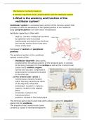
Vestibular system= a somatosensory portion of the nervous system that
provides us with the awareness of the spatial position of our head and
body (proprioception) and self-motion (kinesthesia)
Vestibular apparatus is filled with:
- High K+, low Na+ endolymph secreted
by epithelial cells secreted
continuously and drains from the inner
ear into the venous sinus in the dura
mater of the brain
Composed of central and peripheral
portions
The peripheral portion of the vestibular
system consist of the:
- Vestibular labyrinth: bony cavity
located within the petrous portion of the temporal bone. It consists
of the bony framework for the cochlea as well as the 3 semicircular
canals and 2 otolithic organs
(utricle and saccule). comprised of
proprioceptive components located
in the inner ear:
1) The semicircular canals: 3
membranous channels located
within the bony semicircular ducts
of the labyrinth. Filled with
endolymph. Located in 3 planes:
- superior, located in the sagittal
plane
- horizontal, located in the
transverse plane
- posterior, located in the frontal plane
The terminal part of each canal ends with a dilation called the ampulla
opens into the vestibule
The ampulla of each semicircular canal
contains a cluster of mechanoreceptors=
, crista ampullaris: each crista is composed of special sensory receptors
cells= hair cells. The movements of the fluid (endolymph) stimulate the
hair cells
Crista ampullaris: the cristae respond to changes in the velocity of
rotational movements of the head
each semicircular canal detects when the head moves during rotational
acceleration along its corresponding plane
2) The utricle and saccule (otolithic organs): 2 membranous
cavities that lie in the bony vestibule of the inner ear
Utricle: lies in the posterior part of the
vestibule. On one end it communicates with
the semicircular canals, while on the
opposite end forms a utriculosaccular duct
with the saccule
Saccule: lies anterior to the utricle and is
smaller, besides joining the utriculosaccular
duct, the saccule communicates with the
cochlea by ductus reuniens.
The utricle and saccule contain the cluster of hair
cells as well, but the clusters are called the
macula of the utricle (specialized to detect
movement in the horizontal plane) and the
macula of the saccule (specialized to detect
vertical movement) respond to the stimulation
from the endolymph to detect linear movements of
the head + its position in space while the head is
not moving but the rest of the body is
Each macula is a flat epithelial patch containing
hair cells. Supporting cells surround the macula’s
sensory input from vision, proprioception and the vestibular system
1.What is the anatomy and function of the
vestibular system?
Vestibular system= a somatosensory portion of the nervous system that
provides us with the awareness of the spatial position of our head and
body (proprioception) and self-motion (kinesthesia)
Vestibular apparatus is filled with:
- High K+, low Na+ endolymph secreted
by epithelial cells secreted
continuously and drains from the inner
ear into the venous sinus in the dura
mater of the brain
Composed of central and peripheral
portions
The peripheral portion of the vestibular
system consist of the:
- Vestibular labyrinth: bony cavity
located within the petrous portion of the temporal bone. It consists
of the bony framework for the cochlea as well as the 3 semicircular
canals and 2 otolithic organs
(utricle and saccule). comprised of
proprioceptive components located
in the inner ear:
1) The semicircular canals: 3
membranous channels located
within the bony semicircular ducts
of the labyrinth. Filled with
endolymph. Located in 3 planes:
- superior, located in the sagittal
plane
- horizontal, located in the
transverse plane
- posterior, located in the frontal plane
The terminal part of each canal ends with a dilation called the ampulla
opens into the vestibule
The ampulla of each semicircular canal
contains a cluster of mechanoreceptors=
, crista ampullaris: each crista is composed of special sensory receptors
cells= hair cells. The movements of the fluid (endolymph) stimulate the
hair cells
Crista ampullaris: the cristae respond to changes in the velocity of
rotational movements of the head
each semicircular canal detects when the head moves during rotational
acceleration along its corresponding plane
2) The utricle and saccule (otolithic organs): 2 membranous
cavities that lie in the bony vestibule of the inner ear
Utricle: lies in the posterior part of the
vestibule. On one end it communicates with
the semicircular canals, while on the
opposite end forms a utriculosaccular duct
with the saccule
Saccule: lies anterior to the utricle and is
smaller, besides joining the utriculosaccular
duct, the saccule communicates with the
cochlea by ductus reuniens.
The utricle and saccule contain the cluster of hair
cells as well, but the clusters are called the
macula of the utricle (specialized to detect
movement in the horizontal plane) and the
macula of the saccule (specialized to detect
vertical movement) respond to the stimulation
from the endolymph to detect linear movements of
the head + its position in space while the head is
not moving but the rest of the body is
Each macula is a flat epithelial patch containing
hair cells. Supporting cells surround the macula’s