6BBB0333
L13 & 14 – Engineering Novel Properties: thermal, non-aqueous, and oxidative stability
Evaluating protein stability with respect to temperature, salt and pressure
Currently available thermostable enzymes and their applications
- Looked at enzymes – easy though indirect way of measuring the thermal stability (i.e. if an
enzyme is still active at 90degs, implies it is still folded and active and can measure their
activity to confirm)
- Don’t need sophisticated tool to asses activity
- Process temperature column: temperatures ranging from 30 all the way to 110 (boiling and
beyond)
- Enzymes can adapt to a range of temperatures to operate
- Some processes require high temperatures e.g. baking (need thermostable enzymes)
- E.g. detergents have proteases (break down biomolecules)
Thermostability of protease enzymes from a variety of organisms
,6BBB0333
- Shows same type of protease from different organisms (bacteria)
- Second column – optimal temperature for growth for that particular microorganism (Topt)
- Enzyme taken from that organism (protease) and look at its melting temperature (Tm) or
half-life of enzymatic activity at a given temperature
- Unsurprisingly:
o Organisms that thrive at low temperatures (e.g. B. stearothermophilus), its enzyme
(e.g. Alkaline protease) does not last very long at high temperature
o E.g. D. mucosus Archaelysin Tm is 98 and half-life at 95 is 80 minutes
- Organisms have evolved to live in an ecological niche (extreme temperatures) – enzymes and
proteins needed for life to occur must adapt to conditions
Initial lessons from thermophilic bacteria
Comparing thermophilic vs. mesophilic enzymes
Mesophilic bacteria: optimal temperature of 30-40degs
Thermophilic bacteria: optimal temperature range of 50-60
- Looked at a range of proteins
o Thermolysin
o Phosphoglycerate kinase
o Glyceraldehyde-3-Phosphate dehydrogenase
o Malate dehydrogenase
o Thermitase vs. subtilisin
- Didn’t see anything obvious
- Conclusion:
o Comparing AA sequence and structures revealed no clear mechanism for
thermostability- studying the enzymes from slide 4
o Comparing sequences for thermophiles vs mesophilic showed abundant AA
substitutions
Lys to Arg, Ser to Ala, Gly to Ala/Pro, Asp to Glu
Examples from slide
No theoretical basis for these was immediately apparent
Structural similarities and differences between thermophilic and mesophilic proteins
Similarities
- Both relatively similar
- 40-85% sequence homology
- 3D structures are superimposable
- Same catalytic mechanisms
Differences
- Increased stability of thermozymes attributed to amino acids sequence
,6BBB0333
- Aliphatic index (proportion of protein containing aliphatic (non-aromatic hydrophobic side
chains) AAs) increased in thermophilic proteins (Aliphatic AAs Ala, Val, Ile, Leu etc.)
o Increased contribution to hydrophobic core formation
o Increased three dimensional stability
- Salt bridges- More salt bridges in thermophilic ferredoxin, RNAse H, PGK, malate
dehydrogenase
After further comparisons, some empirical “rules” emerged
- Highlights
o Residue changes from medium to high temperature
o Shift in positively charged residues e.g. from lysine to arginine
o Shift in small residues e.g. serine to alanine
o Glycine frequently replaced by alanine/proline
o Aspartic acid replaced by glutamate
- E.g. Phosphoribosyl anthranilate isomerase
o E.coli (mesophilic): 20 Gly and 5 Pro
o T.maritima (thermophilic): 11 Gly and 11 Pro
- N.B. these were just observations – there were no detailed structures that would allow for
precise comparisons to show what these residues did.
Further Observations
Aliphatic index
- Thermophilic proteins show higher fraction of protein volume occupied by Ala, Val, Ile, Leu
etc.
- When you go from normal to higher temperature you see larger and more hydrophobic
amino acids in the sequence (more of protein volume becoming hydrophobic)
, 6BBB0333
Salt bridges (ion pairs)
- More salt bridges as you increase in temperature
- Additional salt bridges in ferredoxin, RNaseH, PGK, malate dehydrogenase etc. from
thermophiles
Crystallographic B factors
- The B factor is a measure of the average displacement, i.e. the mobility, of each atom in a
structure
- It helps to describe how much atoms in a protein or other biomolecule vibrate or move due
to temperature. A higher B factor indicates more movement or flexibility of the atoms, while
a lower B factor suggests less motion. Essentially, it provides insights into the dynamic
behavior of molecules within a crystal.
- Large B factor – difficult to position atom in the electron density because potentially there is
a bit of movement in the crystal vibration
- It is a measure of local dynamics/flexibility
- Large b factor:
o Atoms are more flexible and dynamic rather than rigid
o Greater uncertainty or variability in the exact positions of atoms in a crystal lattice
due to thermal fluctuations
- Thermostabilty correlates with lower B factor i.e. Stability ∼ Rigidity
- Cold-adapted enzymes show higher B factors and greater flexibility at room temperature
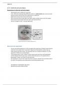
Hyperthermophilic enzyme
First crystal structure: Aldehyde ferredoxin oxidoreductase, 1995
- Topt = 100∘C; Pyrococcus furiosus
- Metallo-protein: Fe4S4 Mo W
- Normal amino-acid composition
- Normal fraction of 2∘ and 3∘ structure
o Usual mixture of alpha helices and beta sheets
- No disulphide bridges
- Small solvent-exposed surface area
o No cavities or crevices
o Small surface area, protein is quite compact
o Volume needed for enzymatic activity squeezed tightly, not extensive reaching out
into space
- Large number of salt bridges
o Iron-sulphur cluster (yellow balls)
L13 & 14 – Engineering Novel Properties: thermal, non-aqueous, and oxidative stability
Evaluating protein stability with respect to temperature, salt and pressure
Currently available thermostable enzymes and their applications
- Looked at enzymes – easy though indirect way of measuring the thermal stability (i.e. if an
enzyme is still active at 90degs, implies it is still folded and active and can measure their
activity to confirm)
- Don’t need sophisticated tool to asses activity
- Process temperature column: temperatures ranging from 30 all the way to 110 (boiling and
beyond)
- Enzymes can adapt to a range of temperatures to operate
- Some processes require high temperatures e.g. baking (need thermostable enzymes)
- E.g. detergents have proteases (break down biomolecules)
Thermostability of protease enzymes from a variety of organisms
,6BBB0333
- Shows same type of protease from different organisms (bacteria)
- Second column – optimal temperature for growth for that particular microorganism (Topt)
- Enzyme taken from that organism (protease) and look at its melting temperature (Tm) or
half-life of enzymatic activity at a given temperature
- Unsurprisingly:
o Organisms that thrive at low temperatures (e.g. B. stearothermophilus), its enzyme
(e.g. Alkaline protease) does not last very long at high temperature
o E.g. D. mucosus Archaelysin Tm is 98 and half-life at 95 is 80 minutes
- Organisms have evolved to live in an ecological niche (extreme temperatures) – enzymes and
proteins needed for life to occur must adapt to conditions
Initial lessons from thermophilic bacteria
Comparing thermophilic vs. mesophilic enzymes
Mesophilic bacteria: optimal temperature of 30-40degs
Thermophilic bacteria: optimal temperature range of 50-60
- Looked at a range of proteins
o Thermolysin
o Phosphoglycerate kinase
o Glyceraldehyde-3-Phosphate dehydrogenase
o Malate dehydrogenase
o Thermitase vs. subtilisin
- Didn’t see anything obvious
- Conclusion:
o Comparing AA sequence and structures revealed no clear mechanism for
thermostability- studying the enzymes from slide 4
o Comparing sequences for thermophiles vs mesophilic showed abundant AA
substitutions
Lys to Arg, Ser to Ala, Gly to Ala/Pro, Asp to Glu
Examples from slide
No theoretical basis for these was immediately apparent
Structural similarities and differences between thermophilic and mesophilic proteins
Similarities
- Both relatively similar
- 40-85% sequence homology
- 3D structures are superimposable
- Same catalytic mechanisms
Differences
- Increased stability of thermozymes attributed to amino acids sequence
,6BBB0333
- Aliphatic index (proportion of protein containing aliphatic (non-aromatic hydrophobic side
chains) AAs) increased in thermophilic proteins (Aliphatic AAs Ala, Val, Ile, Leu etc.)
o Increased contribution to hydrophobic core formation
o Increased three dimensional stability
- Salt bridges- More salt bridges in thermophilic ferredoxin, RNAse H, PGK, malate
dehydrogenase
After further comparisons, some empirical “rules” emerged
- Highlights
o Residue changes from medium to high temperature
o Shift in positively charged residues e.g. from lysine to arginine
o Shift in small residues e.g. serine to alanine
o Glycine frequently replaced by alanine/proline
o Aspartic acid replaced by glutamate
- E.g. Phosphoribosyl anthranilate isomerase
o E.coli (mesophilic): 20 Gly and 5 Pro
o T.maritima (thermophilic): 11 Gly and 11 Pro
- N.B. these were just observations – there were no detailed structures that would allow for
precise comparisons to show what these residues did.
Further Observations
Aliphatic index
- Thermophilic proteins show higher fraction of protein volume occupied by Ala, Val, Ile, Leu
etc.
- When you go from normal to higher temperature you see larger and more hydrophobic
amino acids in the sequence (more of protein volume becoming hydrophobic)
, 6BBB0333
Salt bridges (ion pairs)
- More salt bridges as you increase in temperature
- Additional salt bridges in ferredoxin, RNaseH, PGK, malate dehydrogenase etc. from
thermophiles
Crystallographic B factors
- The B factor is a measure of the average displacement, i.e. the mobility, of each atom in a
structure
- It helps to describe how much atoms in a protein or other biomolecule vibrate or move due
to temperature. A higher B factor indicates more movement or flexibility of the atoms, while
a lower B factor suggests less motion. Essentially, it provides insights into the dynamic
behavior of molecules within a crystal.
- Large B factor – difficult to position atom in the electron density because potentially there is
a bit of movement in the crystal vibration
- It is a measure of local dynamics/flexibility
- Large b factor:
o Atoms are more flexible and dynamic rather than rigid
o Greater uncertainty or variability in the exact positions of atoms in a crystal lattice
due to thermal fluctuations
- Thermostabilty correlates with lower B factor i.e. Stability ∼ Rigidity
- Cold-adapted enzymes show higher B factors and greater flexibility at room temperature
Hyperthermophilic enzyme
First crystal structure: Aldehyde ferredoxin oxidoreductase, 1995
- Topt = 100∘C; Pyrococcus furiosus
- Metallo-protein: Fe4S4 Mo W
- Normal amino-acid composition
- Normal fraction of 2∘ and 3∘ structure
o Usual mixture of alpha helices and beta sheets
- No disulphide bridges
- Small solvent-exposed surface area
o No cavities or crevices
o Small surface area, protein is quite compact
o Volume needed for enzymatic activity squeezed tightly, not extensive reaching out
into space
- Large number of salt bridges
o Iron-sulphur cluster (yellow balls)