Neurocognition week 1 02-09-2024
Lecture 1: Development: The brain and cognition over the life span
This lecture:
Brain structure & anatomy – refresher
- Neurons and other cells in the nervous system.
- Build-up of anatomical structure and naming conventions
Brain development and plasticity
- Natural development over the life span
- Neurogenesis and recovery after damage
- Learning-related plasticity
Development and cognition
- Cognition over the life span
- Clinical intervention strategies
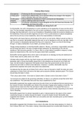
Cells of the brain: neurons and glia cells
Parts you should be able to identify:
- Cell body
- Axon
- Axon hillock
- Dendrites
- Synapse/synaptic cleft
o Pre- and postsynaptic side
o Vesicles and receptors
- Myelin sheath
o Nodes of Ranvier
Neurons, different types, categorized by shape and function:
- Sensory (afferent); carry signals from sensory receptors to the CNS
(Brain+spinal cord).
- Interneurons (stellate, pyramidal, Purkinje); connect neurons within the
CNS and are involved in reflexes and complex processing.
- Motor (efferent); transmit signals from the CNS (brain+spinal cord) to
muscles and glands, facilitating movements and action.
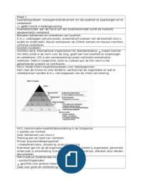
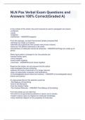
Action potentials
- Thresholded, non-decremental, all-
or-nothing response (it is either
there or not).
- Triggered by summation of
excitatory potentials.
- Driven by varying ion permeability
of cell membrane.
- Propagates along axon an travel
for a meter or more.
- Triggers neurotransmitter release
at axon terminal.
Synapse
- Action potential leads to neurotransmitter release into synaptic cleft
o Some neurons can release more than one type of transmitter
depending on type of stimulation (e.g. low vs high frequency
stimulation)
Acetylcholine
, Dopamine
Norepinephrine
Serotonin
Glutamate
Gamma-aminobutyric acid GABA
- Receptor cells in the postsynaptic membrane can adapt to under or over
use.
o For example, if a particular neurotransmitter is used frequently, the
postsynaptic receptors may become more sensitive or increase in
number, enhancing the neuron's excitability and responsiveness.
- The distribution of synapses connecting to cell influences in excitability.
Glia cells
- Astrocytes
o Blood-brain barrier, structural support.
- Oligodendrocytes
o Responsible for forming myelin for CNS neurons.
- Microglial cells
o Immune cells: fight infections, waste disposal
- Ependymal cells
o Ventricular surface epithelium, create CSF.
- Schwann cells
o Myelin for peripheral neurons (outside the brain)
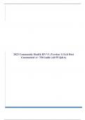
Cortical cell layers
- Different types of neuron are often organized in layers.
- Sensory (input), interneurons (relay) and motor (output) neurons are
groups together.
- Layers are different in different cortical areas, depending on the primary
function.
Each layer has a specific function
In the cortex, neurons are organized into six distinct layers, each serving specific
roles in processing information.
The layers are categorized based on their function:
- Input Layers: These layers primarily receive sensory information from various
sources, including the thalamus, other cortical areas, and the brainstem.
- Relay Layers: Composed of interneurons, these layers facilitate
communication between different cortical areas, relaying information for further
processing.
- Output Layers: These layers send motor commands to the thalamus, other
cortical regions, and subcortical structures, including the brainstem and spinal
cord.
Functional Specialization: Each layer's specific function is influenced by the
type of neurons present and the connections they form. For instance,
certain layers are more densely populated with sensory neurons, while
others may contain more motor neurons.
Interconnectivity: The layers are often interconnected, allowing for
complex processing and integration of information. This interconnectivity is
crucial for the brain's ability to perform higher-level functions such as
decision-making, planning, and motor control.
, White matter tracts: bundles of myelinated axons.
- Connecting neurons throughout the central and peripheral nervous system.
o Association fibers connecting areas within hemisphere.
o Commissural fibers crossing to the other hemisphere, to the same
(homotopic) or a different place (heterotopic).
o Projection fibers connect outward, to subcortical regions, cerebellum
or the spinal cord.
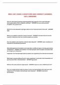
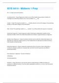
Major component of the CNS
1. Forebrain:
o This includes the largest part of the brain
and includes the cerebral hemispheres,
corpus callosum and subcortical
structures (telencephalon).
o Responsible for higher cognitive
functions, sensory processing and
emotional regulation.
o Telencephalon or cerebrum (cortical
and subcortical):
cortical:
Frontal lobes: movement.
Attention, reward, short-term memory, planning,
impulse control, and more.
Parietal lobes: sensory integration, association
processes, language functions, spatial processing,
sense of touch, some visual processes, and more.
Occipital lobes: mainly primary visual areas.
Temporal lobe: memory, emotion association, primary
auditory areas, some visual processes and more.
Subcortical:
Basal ganglia
o Caudate nucleus
o Putamen
o Globus pallidus
o Subthalamic nucleus
o Substantia nigra
Basal ganglia circuits: multiple circuits in the brain go through the basal
ganglia:
o Motor circuit: organizing voluntary movement through inhibitory and
excitatory pathways.
o Associative circuit; contributing to learning predictive processing
and sequencing.
o Reward circuit; producing pleasure responses, motivational
functions.
Limbic structures: emotional colouring of what you
experience.
o Cingulate -> part of the cortex!
o Hippocampus
o Hypothalamus
o Amygdala
Lecture 1: Development: The brain and cognition over the life span
This lecture:
Brain structure & anatomy – refresher
- Neurons and other cells in the nervous system.
- Build-up of anatomical structure and naming conventions
Brain development and plasticity
- Natural development over the life span
- Neurogenesis and recovery after damage
- Learning-related plasticity
Development and cognition
- Cognition over the life span
- Clinical intervention strategies
Cells of the brain: neurons and glia cells
Parts you should be able to identify:
- Cell body
- Axon
- Axon hillock
- Dendrites
- Synapse/synaptic cleft
o Pre- and postsynaptic side
o Vesicles and receptors
- Myelin sheath
o Nodes of Ranvier
Neurons, different types, categorized by shape and function:
- Sensory (afferent); carry signals from sensory receptors to the CNS
(Brain+spinal cord).
- Interneurons (stellate, pyramidal, Purkinje); connect neurons within the
CNS and are involved in reflexes and complex processing.
- Motor (efferent); transmit signals from the CNS (brain+spinal cord) to
muscles and glands, facilitating movements and action.
Action potentials
- Thresholded, non-decremental, all-
or-nothing response (it is either
there or not).
- Triggered by summation of
excitatory potentials.
- Driven by varying ion permeability
of cell membrane.
- Propagates along axon an travel
for a meter or more.
- Triggers neurotransmitter release
at axon terminal.
Synapse
- Action potential leads to neurotransmitter release into synaptic cleft
o Some neurons can release more than one type of transmitter
depending on type of stimulation (e.g. low vs high frequency
stimulation)
Acetylcholine
, Dopamine
Norepinephrine
Serotonin
Glutamate
Gamma-aminobutyric acid GABA
- Receptor cells in the postsynaptic membrane can adapt to under or over
use.
o For example, if a particular neurotransmitter is used frequently, the
postsynaptic receptors may become more sensitive or increase in
number, enhancing the neuron's excitability and responsiveness.
- The distribution of synapses connecting to cell influences in excitability.
Glia cells
- Astrocytes
o Blood-brain barrier, structural support.
- Oligodendrocytes
o Responsible for forming myelin for CNS neurons.
- Microglial cells
o Immune cells: fight infections, waste disposal
- Ependymal cells
o Ventricular surface epithelium, create CSF.
- Schwann cells
o Myelin for peripheral neurons (outside the brain)
Cortical cell layers
- Different types of neuron are often organized in layers.
- Sensory (input), interneurons (relay) and motor (output) neurons are
groups together.
- Layers are different in different cortical areas, depending on the primary
function.
Each layer has a specific function
In the cortex, neurons are organized into six distinct layers, each serving specific
roles in processing information.
The layers are categorized based on their function:
- Input Layers: These layers primarily receive sensory information from various
sources, including the thalamus, other cortical areas, and the brainstem.
- Relay Layers: Composed of interneurons, these layers facilitate
communication between different cortical areas, relaying information for further
processing.
- Output Layers: These layers send motor commands to the thalamus, other
cortical regions, and subcortical structures, including the brainstem and spinal
cord.
Functional Specialization: Each layer's specific function is influenced by the
type of neurons present and the connections they form. For instance,
certain layers are more densely populated with sensory neurons, while
others may contain more motor neurons.
Interconnectivity: The layers are often interconnected, allowing for
complex processing and integration of information. This interconnectivity is
crucial for the brain's ability to perform higher-level functions such as
decision-making, planning, and motor control.
, White matter tracts: bundles of myelinated axons.
- Connecting neurons throughout the central and peripheral nervous system.
o Association fibers connecting areas within hemisphere.
o Commissural fibers crossing to the other hemisphere, to the same
(homotopic) or a different place (heterotopic).
o Projection fibers connect outward, to subcortical regions, cerebellum
or the spinal cord.
Major component of the CNS
1. Forebrain:
o This includes the largest part of the brain
and includes the cerebral hemispheres,
corpus callosum and subcortical
structures (telencephalon).
o Responsible for higher cognitive
functions, sensory processing and
emotional regulation.
o Telencephalon or cerebrum (cortical
and subcortical):
cortical:
Frontal lobes: movement.
Attention, reward, short-term memory, planning,
impulse control, and more.
Parietal lobes: sensory integration, association
processes, language functions, spatial processing,
sense of touch, some visual processes, and more.
Occipital lobes: mainly primary visual areas.
Temporal lobe: memory, emotion association, primary
auditory areas, some visual processes and more.
Subcortical:
Basal ganglia
o Caudate nucleus
o Putamen
o Globus pallidus
o Subthalamic nucleus
o Substantia nigra
Basal ganglia circuits: multiple circuits in the brain go through the basal
ganglia:
o Motor circuit: organizing voluntary movement through inhibitory and
excitatory pathways.
o Associative circuit; contributing to learning predictive processing
and sequencing.
o Reward circuit; producing pleasure responses, motivational
functions.
Limbic structures: emotional colouring of what you
experience.
o Cingulate -> part of the cortex!
o Hippocampus
o Hypothalamus
o Amygdala