CHAPTER 7
APOPTOSIS
Example questions:
1. DNA damage induces apoptosis via
a. Activation of caspase 9
b. Induction of p53 expression
c. Activation of caspase 3
d. All three of the above mentioned answers are correct
2. Explain how triggering of apoptosis in tumour cells can contribute to new therapies
against cancer, and give an example of a therapy.
Triggering apoptosis in tumour cells can help eliminate cancerous cells by activating
the cell's own programmed death pathways. This prevents the proliferation of
genetically unstable or damaged cells that could lead to tumour growth. One
example of a therapy that utilizes this mechanism is the use of BH3 mimetics, such
as Venetoclax. Venetoclax targets the BCL-2 protein, promoting apoptosis in cancer
cells, particularly in chronic lymphocytic leukaemia (CLL) and other types of cancer.
Definition: Apoptosis is the regulated and orderly destruction of a cell through a genetically
encoded process AKA programmed cell death PCD (this mechanism is intrinsic to the cell).
Apoptosis is organized, neat, and tidy, leaving behind little evidence of the preexisting cell.
The cell undergoing apoptosis is swept clean during phagocytosis by macrophages (neigh-
bouring cells that recognize molecular flags such as phosphatidyl serine).
Function; physiological roles in various organs:
- Developmental morphogenesis: Apoptosis helps shape organs and tissued by removing
unneeded cells, ensuring that structures form correctly
- Control of cell numbers: Apoptosis regulates the number of cells in tissues, preventing
overgrowth and maintaining tissue homeostasis
- Removal of damaged cells: Harmful or damaged beyond repair- cells are removed,
preventing them from accumulating and causing problems (cancer, tissue dysfunction)
- Negative and positive selection of lymphocytes: Eliminates lymphocytes that will attack
the body’s own tissues (negative selection), and supports the survival of beneficial
lymphocytes (positive selection). This helps in the development of a functional and self-
tolerant immune system
,Cytotoxic effect of radio- and chemotherapy
In the context of cancer treatment, apoptosis is a desired outcome. Radiotherapy and
chemotherapy work by inducing apoptosis in cancer cells. By triggering PCD, these
treatments aim to eliminate cancerous cells while minimizing damage to normal tissues.
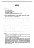
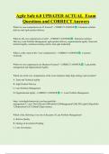
Apoptosis Necrosis
Cell shrinkage Cell swells
Membrane blebbing Leaky membranes
Organelles intact Organelles damaged
Apoptotic antibodies Cell lyses
Chromatin condensation Chromatin
and fragmentation damaged
No inflammation Inflammation
Apoptosis vs. necrosis
Apoptosis is programmed cell death, meaning it is a controlled and planned process. The
cell actively participated in its own destruction, involving a sequence of biochemical events
that lead to cell shrinkage, chromatin condensation, and DNA fragmentation. It maintains
homeostasis and development by eliminating unwanted/harmful cells without causing
inflammation (physiological role).
Necrosis is unplanned and uncontrolled cell death caused by external factors (e.g.,
injury, infection, toxins). The cell swells and bursts, leading to the release of its contents
into the surrounding tissue, which often causes inflammation and damage to nearby cells.
It is typically associated with harmful conditions and can lead to further tissue damage and
inflammation (pathological role).
Apoptosis signalling; induction of apoptosis
Apoptosis can be triggered by various internal and external signals. Here are the key ways
in which apoptosis can be induced:
- Programmed apoptosis: The natural, orderly process by which cells die. It occurs during
normal development and maintenance of tissues
- Loss of growth factors or loss of adhesion: Cells rely on signals from growth factors and
physical contact with other cells or the extracellular matrix to survive. When these
signals are lost, apoptosis can be triggered to remove the affected cells
, - Death receptors of the TNFR family: Tumour necrosis factor receptors are a group of
receptors that, when bound by their specific ligands, can initiate apoptotic signalling
pathways. Examples:
• Fas receptor (CD95)
• TNF-related apoptosis-inducing ligand (TRAIL) receptors
- T- and B-cell antigen receptors: In the immune system, activation of T-cell receptors
(TCRs( or B-cell receptors (BCRs) can lead to apoptosis, especially if these cells are
self-reactive or no longer needed
- Cytotoxic T-lymphocytes: CTLs can induce apoptosis in target cells (e.g., virus-infected
cells, cancer cells) through the release of perforin and granzymes
- DNA damage: Damage to a cell’s DNA (caused by e.g., irradiation or chemotherapy)
can trigger apoptosis. This prevents the propagation of cells with genetic mutations
that could lead to cancer
- Stress conditions: Various stress conditions, including oxidative stress, hypoxia, and
endoplasmic reticulum stress, can activate apoptotic pathways
Mitochondrial changes
Mitochondrial changes play a central role in the intrinsic pathway of apoptosis, including:
- Release of cytochrome c: When the mitochondrial membrane is permeabilized, cyto-
chrome c is released into the cytosol, where it helps to form the apoptosome complex
- Loss of mitochondrial membrane potential: This change is a hallmark of early apoptosis
and leads to the disruption of mitochondrial function
Activation oft he caspase family
Caspases are a family of protease enzymes that play essential roles in apoptosis. Activation
of caspases involve:
- Initiator caspases (2, 8, 9, 10)
• Activated in response to apoptotic signals
• Subsequently activate the executioner caspases
- Executioner caspases (3, 6, 7)
• Execute the death program by cleaving various cellular substrates
Proteolytic cleavage of structural and functional proteins
Once activated, caspases cleave a wide range of cellular proteins, leading to the orderly
dismantling of the cell. This includes:
- Structural proteins: Such as cytoskeletal protein that maintain cell shape and integrity
- Functional proteins: Including enzymes, other proteins critical for cell survival/function
APOPTOSIS
Example questions:
1. DNA damage induces apoptosis via
a. Activation of caspase 9
b. Induction of p53 expression
c. Activation of caspase 3
d. All three of the above mentioned answers are correct
2. Explain how triggering of apoptosis in tumour cells can contribute to new therapies
against cancer, and give an example of a therapy.
Triggering apoptosis in tumour cells can help eliminate cancerous cells by activating
the cell's own programmed death pathways. This prevents the proliferation of
genetically unstable or damaged cells that could lead to tumour growth. One
example of a therapy that utilizes this mechanism is the use of BH3 mimetics, such
as Venetoclax. Venetoclax targets the BCL-2 protein, promoting apoptosis in cancer
cells, particularly in chronic lymphocytic leukaemia (CLL) and other types of cancer.
Definition: Apoptosis is the regulated and orderly destruction of a cell through a genetically
encoded process AKA programmed cell death PCD (this mechanism is intrinsic to the cell).
Apoptosis is organized, neat, and tidy, leaving behind little evidence of the preexisting cell.
The cell undergoing apoptosis is swept clean during phagocytosis by macrophages (neigh-
bouring cells that recognize molecular flags such as phosphatidyl serine).
Function; physiological roles in various organs:
- Developmental morphogenesis: Apoptosis helps shape organs and tissued by removing
unneeded cells, ensuring that structures form correctly
- Control of cell numbers: Apoptosis regulates the number of cells in tissues, preventing
overgrowth and maintaining tissue homeostasis
- Removal of damaged cells: Harmful or damaged beyond repair- cells are removed,
preventing them from accumulating and causing problems (cancer, tissue dysfunction)
- Negative and positive selection of lymphocytes: Eliminates lymphocytes that will attack
the body’s own tissues (negative selection), and supports the survival of beneficial
lymphocytes (positive selection). This helps in the development of a functional and self-
tolerant immune system
,Cytotoxic effect of radio- and chemotherapy
In the context of cancer treatment, apoptosis is a desired outcome. Radiotherapy and
chemotherapy work by inducing apoptosis in cancer cells. By triggering PCD, these
treatments aim to eliminate cancerous cells while minimizing damage to normal tissues.
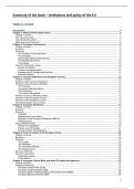
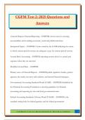
Apoptosis Necrosis
Cell shrinkage Cell swells
Membrane blebbing Leaky membranes
Organelles intact Organelles damaged
Apoptotic antibodies Cell lyses
Chromatin condensation Chromatin
and fragmentation damaged
No inflammation Inflammation
Apoptosis vs. necrosis
Apoptosis is programmed cell death, meaning it is a controlled and planned process. The
cell actively participated in its own destruction, involving a sequence of biochemical events
that lead to cell shrinkage, chromatin condensation, and DNA fragmentation. It maintains
homeostasis and development by eliminating unwanted/harmful cells without causing
inflammation (physiological role).
Necrosis is unplanned and uncontrolled cell death caused by external factors (e.g.,
injury, infection, toxins). The cell swells and bursts, leading to the release of its contents
into the surrounding tissue, which often causes inflammation and damage to nearby cells.
It is typically associated with harmful conditions and can lead to further tissue damage and
inflammation (pathological role).
Apoptosis signalling; induction of apoptosis
Apoptosis can be triggered by various internal and external signals. Here are the key ways
in which apoptosis can be induced:
- Programmed apoptosis: The natural, orderly process by which cells die. It occurs during
normal development and maintenance of tissues
- Loss of growth factors or loss of adhesion: Cells rely on signals from growth factors and
physical contact with other cells or the extracellular matrix to survive. When these
signals are lost, apoptosis can be triggered to remove the affected cells
, - Death receptors of the TNFR family: Tumour necrosis factor receptors are a group of
receptors that, when bound by their specific ligands, can initiate apoptotic signalling
pathways. Examples:
• Fas receptor (CD95)
• TNF-related apoptosis-inducing ligand (TRAIL) receptors
- T- and B-cell antigen receptors: In the immune system, activation of T-cell receptors
(TCRs( or B-cell receptors (BCRs) can lead to apoptosis, especially if these cells are
self-reactive or no longer needed
- Cytotoxic T-lymphocytes: CTLs can induce apoptosis in target cells (e.g., virus-infected
cells, cancer cells) through the release of perforin and granzymes
- DNA damage: Damage to a cell’s DNA (caused by e.g., irradiation or chemotherapy)
can trigger apoptosis. This prevents the propagation of cells with genetic mutations
that could lead to cancer
- Stress conditions: Various stress conditions, including oxidative stress, hypoxia, and
endoplasmic reticulum stress, can activate apoptotic pathways
Mitochondrial changes
Mitochondrial changes play a central role in the intrinsic pathway of apoptosis, including:
- Release of cytochrome c: When the mitochondrial membrane is permeabilized, cyto-
chrome c is released into the cytosol, where it helps to form the apoptosome complex
- Loss of mitochondrial membrane potential: This change is a hallmark of early apoptosis
and leads to the disruption of mitochondrial function
Activation oft he caspase family
Caspases are a family of protease enzymes that play essential roles in apoptosis. Activation
of caspases involve:
- Initiator caspases (2, 8, 9, 10)
• Activated in response to apoptotic signals
• Subsequently activate the executioner caspases
- Executioner caspases (3, 6, 7)
• Execute the death program by cleaving various cellular substrates
Proteolytic cleavage of structural and functional proteins
Once activated, caspases cleave a wide range of cellular proteins, leading to the orderly
dismantling of the cell. This includes:
- Structural proteins: Such as cytoskeletal protein that maintain cell shape and integrity
- Functional proteins: Including enzymes, other proteins critical for cell survival/function