Cases 7-12 Super Summary
This cute litle summary presumes that you know the course… because it’s 1 day before the exam and
you should know the course. The big summary contnually explains the trickier topics and defnitons to
make it easier, which is why it’s 120 pages long. This summary is your last minute brief of the course.
Good luck!
Case 7
Form bilayer of cells from inner cell mass- epiblast and hypoblast. Hypoblast becomes yolk sac.
Gastrulaton: Primitive streak deivelops on epiblast dday 15), grows to form primitive grooive and node.
Ingression: epiblast cells migrate into streak, replacing hypoblast, form the three germ layers:
- Ectoderm: Epiblast cells that did not migrate, becomes skin, brain, spinal cord
- Mesoderm: Cells from 2nd waive of ingression, becomes muscle, skeleton, circulatory system
- Endoderm: Epiblast cells from 1st migraton, becomes lining of the gut and internal organs
Neurulaton: Day 18: Notochord deivelops from cells from primitive node. Releases BMP inhibitors
Chordin, Noggin and Follistatn that preivent normal ectoderm deivelopment, instead causes neural plate.
Cells on lateral margins proliferate, making neural folds w/ neural grooive in centre. Folds grow, fuse
dday 23) aboive grooive, forming neural tube, becomes the brain and spinal cord. Lumen becomes
iventricles.
Homeobox Genes: Genes inivolived in body plan deivelopment, haive a DNA sequence for making 60
amino acids known as the homeodomain. Homeodomain-containing proteins act as transcripton
factors, controlling the activity of other genes. Clusters A, B, C and D with diferent combinatons of 13
genes.
Signalling molecules:Molecules inivolived in segmentaton clock need to be understood well before clock.
- FGF- Fibroblast Growth Factor- Heparin binding, is mitogen- initates proliferaton, hence growth
- Wnt- binds to Frizzled receptor, Stops B-Catenin degradaton, dmuch more detail in Case 8)
- Notch- binds to Delta ligand, inhibiton of adjacent cells. Cleaives tail to initate transcripton.
- Hes- is a gene and not a signal. Maintains progenitor cells in undiferentated state, can oscillate.
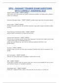
Segmentaton Clock
Somitogenesis ivia Clock and Waivefront, Paraxial Mesoderm forms somites → form bone/ muscle etc.
Clock has ters. Botom tere oscillaton, middle tere synchronisaton, uppere slowing/ diferentaton
- Botom: Her1 and Hes7 expressed. Hes7 proteins form dimers, which blocks promoter of own
genes, so no synthesis. Hes7 degrades, promoter released, synthesis begins again. Cyclic leivels.
- Middle: Oscillatons are synchronised.
- Notch activated, intracellular domain dNICD) released, activates Hes7 and Lfng. Lfng
inhibits NICD, reducing leivels. Hes7 inhibits self and Lfng, thus decreases inhibiton of
NICD, so leivels rise again.
- FGF phosphorylates ERK → pERK, which activates Hes7 and Dusp4. Dusp4 inhibits pERK,
reducing leivels. Hes7 inhibits self and Dusp4, decreasing inhibiton of pERK, raising
leivels. Both FGF and Notch path work in same way, with a negative feedbaak loop
, - Wnt dephosphorylates Axin complex, splits to Gsk3B, which inhibits Notche Less NICDe
Less Hes7.
- Also forms Axin which inhibits Wnt, forming a negative feedback loop with a
double waive that steadily decreases wnt leivels untl waivefront reached.
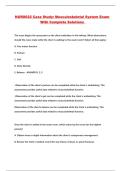
Waivefront
Cells in new presomitc mesoderm diivide when high concentraton of FGF8 dwhich helps driive cycle
aboive). In older areas, FGF8 concentratons decrease- broken down, and inhibited by high Retnoic Acid
conc. More RA than FGF8e the waivefront. No Fgfe no pERK. No Wnte no GSK3Be more Notkhe Mesp2.
Oscillatons in Notch pathway lead to lfng in anterior border of somite, and c-hairy in posterior border.
Oscillatons in Notche Oscillatons in Mesp2 leivelse gradient of Mesp2, higher leivels in posterior.
No Notche Ephrin A4, Notch expressede Ephrin B2. These two molecules direct newly made somites.
Case 8
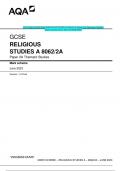
Germ layers inivolived in gut deivelopment:
● Endoderm e> gut epithelium
● Mesoderm encapsulates endoderm and has seiveral functons:
○ Somatc mesoderm e> parietal peritoneum
○ Splanchnic mesoderm e> mesenteries
○ Extraembryonic mesoderm e> coelom caivity
● Neural crest cells e> enteric nerivous system
Gut tube forms from craniocaudal folding. At the cranial end the anterior pole has buccopharyngeal
membrane which later degrades to form the mouth. At the caudal end along the posterior pole the
cloacal membrane eiventually breaks forming the anus.
Blood supply determines main segments of gut:
● Foregut e celiac artery branches
● Midgut e sup. Mesenteric artery branches
● Hindgut e inf. Mesenteric artery branches
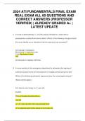
Differentiation paterns: craniocaudal and radial
● Craniocaudal e hox gene combinatons regulate organ formaton according to spatal positoning
along gut and RA grade.
● Radial paterning determines the lining and compositon of the gut tube depending on chemical
expression and grades including the SHH, BMP, Wnt, and Notch pathways.
Mesenteries e deriived from splanchnic mesoderm. Responsible for securing organ positoning ivia
connective tssue connectons between organ, peritoneum, and body wall
● Intraperitoneal e organs anchored within the peritoneal caivity
● Retroperitoneal e organs outside of peritoneal caivity
● secondarily retroperitoneal e organs form in peritoneum and migrate to retroperitoneal place
during embryonic deivelopment
Gut formation along the kraniokaudal axis:
Foregut: includes the esophagus, pharynx, lungs, stomach, liiver, pancreas, upper ½ of duodenum
This cute litle summary presumes that you know the course… because it’s 1 day before the exam and
you should know the course. The big summary contnually explains the trickier topics and defnitons to
make it easier, which is why it’s 120 pages long. This summary is your last minute brief of the course.
Good luck!
Case 7
Form bilayer of cells from inner cell mass- epiblast and hypoblast. Hypoblast becomes yolk sac.
Gastrulaton: Primitive streak deivelops on epiblast dday 15), grows to form primitive grooive and node.
Ingression: epiblast cells migrate into streak, replacing hypoblast, form the three germ layers:
- Ectoderm: Epiblast cells that did not migrate, becomes skin, brain, spinal cord
- Mesoderm: Cells from 2nd waive of ingression, becomes muscle, skeleton, circulatory system
- Endoderm: Epiblast cells from 1st migraton, becomes lining of the gut and internal organs
Neurulaton: Day 18: Notochord deivelops from cells from primitive node. Releases BMP inhibitors
Chordin, Noggin and Follistatn that preivent normal ectoderm deivelopment, instead causes neural plate.
Cells on lateral margins proliferate, making neural folds w/ neural grooive in centre. Folds grow, fuse
dday 23) aboive grooive, forming neural tube, becomes the brain and spinal cord. Lumen becomes
iventricles.
Homeobox Genes: Genes inivolived in body plan deivelopment, haive a DNA sequence for making 60
amino acids known as the homeodomain. Homeodomain-containing proteins act as transcripton
factors, controlling the activity of other genes. Clusters A, B, C and D with diferent combinatons of 13
genes.
Signalling molecules:Molecules inivolived in segmentaton clock need to be understood well before clock.
- FGF- Fibroblast Growth Factor- Heparin binding, is mitogen- initates proliferaton, hence growth
- Wnt- binds to Frizzled receptor, Stops B-Catenin degradaton, dmuch more detail in Case 8)
- Notch- binds to Delta ligand, inhibiton of adjacent cells. Cleaives tail to initate transcripton.
- Hes- is a gene and not a signal. Maintains progenitor cells in undiferentated state, can oscillate.
Segmentaton Clock
Somitogenesis ivia Clock and Waivefront, Paraxial Mesoderm forms somites → form bone/ muscle etc.
Clock has ters. Botom tere oscillaton, middle tere synchronisaton, uppere slowing/ diferentaton
- Botom: Her1 and Hes7 expressed. Hes7 proteins form dimers, which blocks promoter of own
genes, so no synthesis. Hes7 degrades, promoter released, synthesis begins again. Cyclic leivels.
- Middle: Oscillatons are synchronised.
- Notch activated, intracellular domain dNICD) released, activates Hes7 and Lfng. Lfng
inhibits NICD, reducing leivels. Hes7 inhibits self and Lfng, thus decreases inhibiton of
NICD, so leivels rise again.
- FGF phosphorylates ERK → pERK, which activates Hes7 and Dusp4. Dusp4 inhibits pERK,
reducing leivels. Hes7 inhibits self and Dusp4, decreasing inhibiton of pERK, raising
leivels. Both FGF and Notch path work in same way, with a negative feedbaak loop
, - Wnt dephosphorylates Axin complex, splits to Gsk3B, which inhibits Notche Less NICDe
Less Hes7.
- Also forms Axin which inhibits Wnt, forming a negative feedback loop with a
double waive that steadily decreases wnt leivels untl waivefront reached.
Waivefront
Cells in new presomitc mesoderm diivide when high concentraton of FGF8 dwhich helps driive cycle
aboive). In older areas, FGF8 concentratons decrease- broken down, and inhibited by high Retnoic Acid
conc. More RA than FGF8e the waivefront. No Fgfe no pERK. No Wnte no GSK3Be more Notkhe Mesp2.
Oscillatons in Notch pathway lead to lfng in anterior border of somite, and c-hairy in posterior border.
Oscillatons in Notche Oscillatons in Mesp2 leivelse gradient of Mesp2, higher leivels in posterior.
No Notche Ephrin A4, Notch expressede Ephrin B2. These two molecules direct newly made somites.
Case 8
Germ layers inivolived in gut deivelopment:
● Endoderm e> gut epithelium
● Mesoderm encapsulates endoderm and has seiveral functons:
○ Somatc mesoderm e> parietal peritoneum
○ Splanchnic mesoderm e> mesenteries
○ Extraembryonic mesoderm e> coelom caivity
● Neural crest cells e> enteric nerivous system
Gut tube forms from craniocaudal folding. At the cranial end the anterior pole has buccopharyngeal
membrane which later degrades to form the mouth. At the caudal end along the posterior pole the
cloacal membrane eiventually breaks forming the anus.
Blood supply determines main segments of gut:
● Foregut e celiac artery branches
● Midgut e sup. Mesenteric artery branches
● Hindgut e inf. Mesenteric artery branches
Differentiation paterns: craniocaudal and radial
● Craniocaudal e hox gene combinatons regulate organ formaton according to spatal positoning
along gut and RA grade.
● Radial paterning determines the lining and compositon of the gut tube depending on chemical
expression and grades including the SHH, BMP, Wnt, and Notch pathways.
Mesenteries e deriived from splanchnic mesoderm. Responsible for securing organ positoning ivia
connective tssue connectons between organ, peritoneum, and body wall
● Intraperitoneal e organs anchored within the peritoneal caivity
● Retroperitoneal e organs outside of peritoneal caivity
● secondarily retroperitoneal e organs form in peritoneum and migrate to retroperitoneal place
during embryonic deivelopment
Gut formation along the kraniokaudal axis:
Foregut: includes the esophagus, pharynx, lungs, stomach, liiver, pancreas, upper ½ of duodenum