TASK 3: EEG & ERP AS DEPENDENT
VARIABLES
NEURAL ORIGINS OF EEG
SINGLE-CELL RECORDINGS IN ANIMALS
Enabled researchers to describe response characteristics of individual elements
How does it work?
Thin electrode inserted into animal’s brain
When electrode is in vicinity of neuronal membrane, changes in electrical activity
can be measured
Done extracellularly (electrode outside the neuron)
Typical experiment
Recordings obtained from series of cells in target area of interest
Functional map can describe similarities & differences between neurons in cortical
region
Visual areas
Single cell not responsive to all visual stimuli
Receptive fields – a limited region of space to which a specific neuron / cell responds
Neighbouring cells have partially overlapping receptive fields
Retinotopic – topographic representation in vision
Multiunit recording – looking at pattern of activity over a group of neurons (today >400
cells simultaneously)
SINGLE-CELL RECORDINGS IN HUMANS
Only when surgical procedure is required to treat patient (e.g., for epilepsy)
Commonly placed in medial temporal lobe (MTL)
MTL neurons respond selective to specific familiar images
ELECTROCORTOGRAM (ECOC)
Similar to EEG BUT electrodes placed directly on surface of the brain (outside / beneath
dura)
Appropriate only for people undergoing neurosurgical treatment
Electrodes measure electrical signals before they pass through the scalp & skull
less distortion
Excellent spatial & temporal resolution
Used to stimulate the brain & to map and localise cortical & subcortical neurologic
functions
Limitation – experimental question often dictated by location of the ECoC grid
, THE NEURONAL SOURCE OF EEG
EEG arises from synchronised synaptic activity in populations of cortical neurons
Excitation of postsynaptic neurons Dipole – extracellular voltage near neural
dendrites that is more negative than elsewhere along the neurons
Source – region of positive charge separated from a region of negative charge
by some distance
Sink – region of negative charge
Electrodes detect sum of positive & negative charges in their vicinity
Can only detect dipoles if electrode is closer to positive OR negative end of dipole
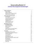
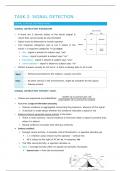
Dipoles 2 major types of dipoles
Radial dipoles – oriented
perpendicular to the
surface (a)
Tangential dipoles – oriented parallel to
the scalp surface (b)
Dipoles have positive & negative side produce both positive & negative
deflection at different regions of the scalp
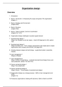
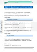
Dipoles from multiple neurons in a region will sum together
In order to achieve a measurable (non-zero) signal, neurons
must be both
Arranged in parallel fashion (a)
Signals can sum to form larger signal
Any other configuration (c) – individual dipoles’ positive &
negative ends will cancel each other out
Synchronously active
Yields a net charge on the scalp-facing side of the dipole
sheet (a) rather than charges cancelling each other out
(b)
Signal large enough to be measured
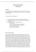
Polarity measured at scalp also depends on particular orientation of dipole
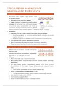
Excitatory postsynaptic potential (EPSP)
EPSP triggered at dendritic synapse the local extracellular fluid becomes more
negative compared to intracellular fluid due to depolarising current in the
neuron
Current flows elsewhere along neuron more distributed extracellular
negativity
VARIABLES
NEURAL ORIGINS OF EEG
SINGLE-CELL RECORDINGS IN ANIMALS
Enabled researchers to describe response characteristics of individual elements
How does it work?
Thin electrode inserted into animal’s brain
When electrode is in vicinity of neuronal membrane, changes in electrical activity
can be measured
Done extracellularly (electrode outside the neuron)
Typical experiment
Recordings obtained from series of cells in target area of interest
Functional map can describe similarities & differences between neurons in cortical
region
Visual areas
Single cell not responsive to all visual stimuli
Receptive fields – a limited region of space to which a specific neuron / cell responds
Neighbouring cells have partially overlapping receptive fields
Retinotopic – topographic representation in vision
Multiunit recording – looking at pattern of activity over a group of neurons (today >400
cells simultaneously)
SINGLE-CELL RECORDINGS IN HUMANS
Only when surgical procedure is required to treat patient (e.g., for epilepsy)
Commonly placed in medial temporal lobe (MTL)
MTL neurons respond selective to specific familiar images
ELECTROCORTOGRAM (ECOC)
Similar to EEG BUT electrodes placed directly on surface of the brain (outside / beneath
dura)
Appropriate only for people undergoing neurosurgical treatment
Electrodes measure electrical signals before they pass through the scalp & skull
less distortion
Excellent spatial & temporal resolution
Used to stimulate the brain & to map and localise cortical & subcortical neurologic
functions
Limitation – experimental question often dictated by location of the ECoC grid
, THE NEURONAL SOURCE OF EEG
EEG arises from synchronised synaptic activity in populations of cortical neurons
Excitation of postsynaptic neurons Dipole – extracellular voltage near neural
dendrites that is more negative than elsewhere along the neurons
Source – region of positive charge separated from a region of negative charge
by some distance
Sink – region of negative charge
Electrodes detect sum of positive & negative charges in their vicinity
Can only detect dipoles if electrode is closer to positive OR negative end of dipole
Dipoles 2 major types of dipoles
Radial dipoles – oriented
perpendicular to the
surface (a)
Tangential dipoles – oriented parallel to
the scalp surface (b)
Dipoles have positive & negative side produce both positive & negative
deflection at different regions of the scalp
Dipoles from multiple neurons in a region will sum together
In order to achieve a measurable (non-zero) signal, neurons
must be both
Arranged in parallel fashion (a)
Signals can sum to form larger signal
Any other configuration (c) – individual dipoles’ positive &
negative ends will cancel each other out
Synchronously active
Yields a net charge on the scalp-facing side of the dipole
sheet (a) rather than charges cancelling each other out
(b)
Signal large enough to be measured
Polarity measured at scalp also depends on particular orientation of dipole
Excitatory postsynaptic potential (EPSP)
EPSP triggered at dendritic synapse the local extracellular fluid becomes more
negative compared to intracellular fluid due to depolarising current in the
neuron
Current flows elsewhere along neuron more distributed extracellular
negativity