GGZ2025 Neuropsychological Disorders vvanbeek
TASK 1 – NEUROANATOMY AND BRAIN IMAGING
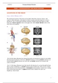
LOCATIONS IN THE BRAIN
Source: Kolb & Whishaw (2015)
The anatomical locations of the brain are described within three reference frames; with
respect to other body parts, with respect to relative location, and with respect to a viewer’s
perspective. Part A of the figure describes structures oriented in the head relative to other
body parts. Dorsal (superior) refers to the upper side, while ventral (inferior) refers to the
lower side of an organism.
-Part B of the figure illustrates how human brain parts are described in relation to one another
from the frame of reference of the fase. Anterior (frontal) is in front, posterior structures are
located behind, lateral structures are at the side, and medial structures are located at the
centre or between.
-Part C of the figure illustrates terms that describe the direction of a section through the brain
from a viewer’s perspective. A coronal section reveals a frontal view, and is cut in a vertical
plane from the crown of the head down. A horizontal section produces a dorsal view, looking
down on the brain from above. A sagittal section reveals a medial view and is cut
lengthways, front to back.
1
, GGZ2025 Neuropsychological Disorders vvanbeek
The nervous system is also symmetrical. Structures that lie on the
same side are ipsilateral, and if they lie on the opposite sides, they are
contralateral to each other. If one lies in each hemisphere, the
structures are bilateral.
Structures close to one another are proximal; those far from another
are distal. Any movement toward a brain structure is afferent,
whereas movement away from is efferent. So, motor pathways leading
to the body from the brain are efferent.
Nervous Systems structure and function
The central nervous system (CNS) consists of the brain and the spinal cord, both encased in
bone. The peripheral nervous system (PNS) has two divisions:
1) The somatic nervous systems (SNS) consists of two sets of inputs and outputs to the
CNS. These are spinal and cranial nerves to and from sensory organs and muscles,
joints and skin. The SNS transmits incoming sensory information and the position and
movement of body parts to the CNS, and produces movements in response.
2) The autonomic nervous system (ANS) controls the functioning of the body’s internal
organs to ‘rest and digest’ through the parasympathetic nerves (calming) or to ‘fight
and flee’ trough the sympathetic nerves (arousing).
Support and protection
The brain and spinal cord are supported and protected from injury and infection in four ways.
(1) The brain is enclosed in the skull, and the spinal cord is encased in a series of interlocking
bony vertebrae. Thus, the CNS lies within bony encasement, whereas the SNS and ANS lie
outside them. Lacking bony protection, the PNS divisions are more vulnerable to injury, but
they can renew themselves after injury. Self-repair is much more limited within the CNS.
(2) Within the bony case enclosing the CNS
is a triple layered set of membranes, the
meninges. The outer dura mater is a though
double layer of tissue enclosing the brain in a
kind of loose sack. The middle arachnoid
membrane is a very thin sheet of delicate
tissue that follows the brain’s contours. The
inner pia matter is a moderately though
tissue that clings to the brain’s surface.
2
, GGZ2025 Neuropsychological Disorders vvanbeek
(3) The brain and spinal cord are cushioned from shock and sudden pressure by the
cerebrospinal fluid (CSF), which circulates through the brain’s four ventricles, the spinal
column and within the subarachnoid space in the brain’s meninges. CSF is continually being
made and drained off into the circulatory system through connecting channels among the
ventricles. If the outflow in these channels is blocked, severe intellectual impairments and
even death can result from built-up CSF pressure.
(4) The blood-brain barrier protects the brain and spinal cords by limiting movement of
chemicals from the rest of the body into the CNS. Glial cells called astroglia stimulate the
cells of capillaries to form tight junctions with one another, thus preventing blood-borne
substances from crossing into the CNS.
Blood supply
The brain receives its blood supply from two internal carotid arteries and two vertebral
arteries that course up each side of the neck. The four arteries connect at the base of the brain,
where they enter the skull. They branch off into several smaller arteries that irrigate the
brainstem and cerebellum and give rise to three cerebral arteries that irrigate the forebrain.
-The anterior cerebral artery (ACA)
irrigates the medial and dorsal parts of
the cortex.
-The middle cerebral artery (MCA)
irrigates the lateral surface of the cortex.
-The posterior cerebral artery (PCA)
irrigates its ventral and posterior
surfaces.
THE BRAINSTEM
The brainstem begins where the spinal cord enters the skull and extends upward into the
lower areas of the forebrain. A distinctive part of the brainstem comprises the many cranial-
nerve nuclei that converge at its core and send their axons to the head muscles. The brainstem
core consists of those cranial-nerve nuclei and other nuclei that mediate a variety of
regulatory functions.
The brainstem consists of three main regions:
the diencephalon, the midbrain and the
hindbrain.
3
TASK 1 – NEUROANATOMY AND BRAIN IMAGING
LOCATIONS IN THE BRAIN
Source: Kolb & Whishaw (2015)
The anatomical locations of the brain are described within three reference frames; with
respect to other body parts, with respect to relative location, and with respect to a viewer’s
perspective. Part A of the figure describes structures oriented in the head relative to other
body parts. Dorsal (superior) refers to the upper side, while ventral (inferior) refers to the
lower side of an organism.
-Part B of the figure illustrates how human brain parts are described in relation to one another
from the frame of reference of the fase. Anterior (frontal) is in front, posterior structures are
located behind, lateral structures are at the side, and medial structures are located at the
centre or between.
-Part C of the figure illustrates terms that describe the direction of a section through the brain
from a viewer’s perspective. A coronal section reveals a frontal view, and is cut in a vertical
plane from the crown of the head down. A horizontal section produces a dorsal view, looking
down on the brain from above. A sagittal section reveals a medial view and is cut
lengthways, front to back.
1
, GGZ2025 Neuropsychological Disorders vvanbeek
The nervous system is also symmetrical. Structures that lie on the
same side are ipsilateral, and if they lie on the opposite sides, they are
contralateral to each other. If one lies in each hemisphere, the
structures are bilateral.
Structures close to one another are proximal; those far from another
are distal. Any movement toward a brain structure is afferent,
whereas movement away from is efferent. So, motor pathways leading
to the body from the brain are efferent.
Nervous Systems structure and function
The central nervous system (CNS) consists of the brain and the spinal cord, both encased in
bone. The peripheral nervous system (PNS) has two divisions:
1) The somatic nervous systems (SNS) consists of two sets of inputs and outputs to the
CNS. These are spinal and cranial nerves to and from sensory organs and muscles,
joints and skin. The SNS transmits incoming sensory information and the position and
movement of body parts to the CNS, and produces movements in response.
2) The autonomic nervous system (ANS) controls the functioning of the body’s internal
organs to ‘rest and digest’ through the parasympathetic nerves (calming) or to ‘fight
and flee’ trough the sympathetic nerves (arousing).
Support and protection
The brain and spinal cord are supported and protected from injury and infection in four ways.
(1) The brain is enclosed in the skull, and the spinal cord is encased in a series of interlocking
bony vertebrae. Thus, the CNS lies within bony encasement, whereas the SNS and ANS lie
outside them. Lacking bony protection, the PNS divisions are more vulnerable to injury, but
they can renew themselves after injury. Self-repair is much more limited within the CNS.
(2) Within the bony case enclosing the CNS
is a triple layered set of membranes, the
meninges. The outer dura mater is a though
double layer of tissue enclosing the brain in a
kind of loose sack. The middle arachnoid
membrane is a very thin sheet of delicate
tissue that follows the brain’s contours. The
inner pia matter is a moderately though
tissue that clings to the brain’s surface.
2
, GGZ2025 Neuropsychological Disorders vvanbeek
(3) The brain and spinal cord are cushioned from shock and sudden pressure by the
cerebrospinal fluid (CSF), which circulates through the brain’s four ventricles, the spinal
column and within the subarachnoid space in the brain’s meninges. CSF is continually being
made and drained off into the circulatory system through connecting channels among the
ventricles. If the outflow in these channels is blocked, severe intellectual impairments and
even death can result from built-up CSF pressure.
(4) The blood-brain barrier protects the brain and spinal cords by limiting movement of
chemicals from the rest of the body into the CNS. Glial cells called astroglia stimulate the
cells of capillaries to form tight junctions with one another, thus preventing blood-borne
substances from crossing into the CNS.
Blood supply
The brain receives its blood supply from two internal carotid arteries and two vertebral
arteries that course up each side of the neck. The four arteries connect at the base of the brain,
where they enter the skull. They branch off into several smaller arteries that irrigate the
brainstem and cerebellum and give rise to three cerebral arteries that irrigate the forebrain.
-The anterior cerebral artery (ACA)
irrigates the medial and dorsal parts of
the cortex.
-The middle cerebral artery (MCA)
irrigates the lateral surface of the cortex.
-The posterior cerebral artery (PCA)
irrigates its ventral and posterior
surfaces.
THE BRAINSTEM
The brainstem begins where the spinal cord enters the skull and extends upward into the
lower areas of the forebrain. A distinctive part of the brainstem comprises the many cranial-
nerve nuclei that converge at its core and send their axons to the head muscles. The brainstem
core consists of those cranial-nerve nuclei and other nuclei that mediate a variety of
regulatory functions.
The brainstem consists of three main regions:
the diencephalon, the midbrain and the
hindbrain.
3