Pathophysiology
Atherosclerosis: Vascular Biology and Nuclear receptors 09/10/2017
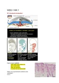
Atherosclerosis (Slagader verkalking): underlying pathology of myocardial and cerebral infarction.
High LDL, high cholesterol is one of the causes. LDL is modified by reactive oxygen species,
endothelial cells become activated, making monocytes bind to endothelial cells. These monocytes
become macrophages inside the endothelium. Macrophages take up the mLDL, secrete cytokines and
growth factors and become foam cells that get stuck in the wall. Growth factors cause smooth muscle
cells to migrate to the endothelium and grow. Multifactorial disease. Lesion formation by: EC,
monocytes/Mf, SMC, Tcell.
Causes: smoking, diet, lack of exercise.
Risk factors: High LDL (statins), low HDL, hypertension, diabetes (insulin)
Genetic components: Familiar hypercholesterolemia (LDL receptor mutation), Tangiers
disease (ABCA1 transporter -> low HDL levels). Combination of different genes.
Treatment: stent to open up the vessel. 1 in 4 patients develop in-stent restenosis.
Key players
SMC: relaxation and contraction of vessel, migrates to endothelium in coagulation, less
contraction.
Endothelial cells: border between blood and tissue, when opened up the coagulation
cascade is started.
Macrophages: take up mLDL, secrete cytokines + Growth factors. Become foam cells
T cells: macrophages present antigens, the T cells respond to that, as a result more cytokines
are synthesized.
Acute myocardial infarction: plaque rupture, when it is obstructing blood flow. When there is a
rupture in the plaques, the endothelial layer will be opened up, coagulation happens, blood clot.
Stable plaque: SMC give sturdiness to the plaque, making it rupture less easily.
Vulnerable plaque: less SMC, more lipids in the plaque.
In-stent restenosis: Stent used as treatment for Atherosclerosis can cause in-stent restenosis. This is a
vascular pathology caused by rapid growth of vascular smooth muscle cells in the stented artery
segment. Predominantly has smooth muscle cells, smooth muscle cell proliferation. To discover the
genes involved in this disease, SNPs involved in muscle cell proliferation were looked for.
P27kip1: SNP gene in in-stent restenosis patients. SMC proliferation is inhibited by P27kip1.
Associated with in-stent restenosis. Inhibitor of the cell cycle in S-phase, check point.
SNP C variant: has less p27 expression than the A variant. Less SMC proliferation
inhibition.
A variant: Higher SMC proliferation inhibition. Has a low chance of developing in-
stent restenosis but an increased risk of acute myocardial infarction. Because it has
lower SMC proliferation, so muscle cells will form less quickly, preventing in-stent
muscle formation. In myocardial infarction, the smooth muscle cells form a stable
plaque, so without the high SMC proliferation, the plaque is more vulnerable.
Nuclear hormone receptor NUR77: small ligand dependent activation, suitable for drug targets.
Transcription factor that activates M1/M2 macrophages. NUR77 knockout mice had more
arteriosclerotic lesions and express more SDF-1alpha. Nur77 has a protective function against
Atherosclerosis, activators could be used in treatment. NUR77 protects endothelium, inhibits
macrophage activation and prevents SMC proliferation.
Atherosclerotic mouse: LDLR or ApoE knockout mouse is given a bone marrow transplantation of a
Nur77 knockout mouse. Normal mice don’t develop atherosclerosis because they have high HDL and
very low LDL. Knockout is therefore needed to mimic atherosclerosis.
1
,Heart failure and Nuclear receptors 09/10/2017
Heart failure: contractility of the left ventricle decreases, meaning that the heart is unable to pump
enough blood to meet the body’s needs for blood and oxygen. Heart failure has different underlying
pathologies, specific treatment is therefore hard. Involves multiple cell types and organ systems.
Myocardial infarction: Ischemia (blood cannot reach the heart) -> cardiomyocytes die -> decreased
contractility and rupture -> decreased cardiac output -> death. Immune cells infiltrate the heart after
infarction, these immune cells are positive for NUR77. Influx of Ly6Chigh cells.
NUR77: high expression in the healthy and diseased heart. NUR77 is needed to transform Ly6Chigh
monocytes into Ly6Clow. Knockout mice only have the Ly6Chigh anti-inflammatory monocytes. In a
mouse model of myocardial infarction, the left coronary artery was closed up in Nur77-KO and wild-
type mice. FACS analysis -> Wild type had Ly6Chigh and low, KO had only Ly6Chigh.
Ly6Chigh: pro-inflammatory, cause cell death and phagocytose.
Ly6Clow: anti-inflammatory, cleanup of debris, formation of protective scar.
Cardiac compensation: heart tries to compensate for the heart failure by changing shape or increase
contractility.
Pathological hypertrophy: stimulus causes to contract more, to pump enough blood. When
this grows too thick, this is dangerous when blood is low because the cavity is small. Caused
by fetal gene expression, which is not healthy in adults. NUR77 protects against
cardiomyocyte hypertrophy because it lowers calcium levels. Researched using siRNA
mediated knockdown of NUR77.
Calcium: is the second messenger for contraction of cardiomyocytes. The calcium is
released from the SR when calcium enters the cytoplasm. Calcium binding to
Troponin causes heart contractions. Ca2+ also activates kinases that activate
transcription factors for hypertrophy gene expression. NUR77 decreases calcium
levels, which explains the lower rate of hypertrophy.
Cardiac dilation & heart failure: heart becomes bigger, so the heart muscle becomes smaller.
Long cardiomyocytes. Can’t empty itself all the way, decreased cardiac output. Myocytes die.
Via sympathetic nervous system (fight/flight): increases heart contractility. Adrenalin binds
to beta adrenergic receptors on the heart, which increases calcium (Beta blockers block this
effect, to calm you down). Cofactors are also released, Neuropeptide Y can bind to receptors
on the heart, which has the same effect as adrenalin but can also increase the effect of
adrenalin. NPY causes hypertrophy.
NUR77: inhibits Neuropeptide Y in macrophages, but also in adrenal cells! -> less mRNA and protein.
NUR77 knockouts had higher mRNA and protein levels for NPY. NUR77 inhibits cardiomyocyte
hypertrophy and cardiac fibrosis in vivo via circulating NPY (paracrine). Hypertrophy is higher in
NUR77 knockouts than in NUR77-KO with antagonist for NPY receptor, this shows that NUR77
decreases hypertrophy.
- promotes repair monocyte differentiation after Miocardial Infarction,
- reduces calcium and hypertrophy in cardiomyocytes,
- reduces cardiac fibrosis
- inhibits systemic expression of pro-hypertrophic NPY.
Isoproterenol: stable form of adrenalin, to stimulate hypertrophy in mice. Via pump by osmosis. NPY
receptor antagonist + isoproterenol is used as a test to see difference from only isoproterenol. When
NPY receptor antagonist was added, the cardiomyocyte size was smaller than with only isoproterenol
in NUR77KO mice. In wild type and WT + antagonist, cardiomyocytes were smaller.
2
, Cholesterol 09/10/2017
Atherosclerosis: can happen in all organs, arteries are blocked and parts of the organ will die. When
cholesterol is too high, the body will release it to get rid of it, clogging the arteries.
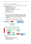
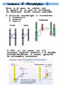
Cholesterol: is an essential lipid, insoluble in water and can’t be broken down. Its level must be
maintained very narrowly, dysregulation causes atherosclerosis. Cholesterol synthesis is very intense
energetically, so LDL receptor upregulation is easier. Cholesterol regulates its own synthesis by
controlling INSIG, SCAP and SREPB2 complex, but also by non-transcriptional mechanisms. This
regulation is subjected to multiple feedback mechanisms, like microRNA 33 inhibition of cholesterol
efflux, cholesterol dependent degradation of SQLE by MARCH6 and the degradation of the LDLR by
IDOL.
Low cholesterol: SREBP causes more cholesterol synthesis and increases uptake. SREBP
active, transport of complex to Golgi. SREBP downregulates ABCA1 with microRNA 33, cellular
cholesterol goes up because it cannot be transported out anymore. SREBP-2 activates the
entire pathway for cholesterol synthesis. Regulation happens in the ER. When cholesterol is
low, INSIG-SCAP-SREPB2 complex is broken down, and the transcription factor SREBP2
becomes active. INSIG, the inhibitor dissociates. SCAP measures cholesterol.
High cholesterol: Nuclear receptors LXRs are activated by oxysterol (cholesterol metabolites)
ligands, this activates a genetic program that promotes cholesterol efflux. LXR induces IDOL,
which ubiquinates LDLreceptors for degradation. INSIG-SCAP-SREPB2 complex is held
together, so the SREBP TF can’t become active. Cholesterol activates MARCH6, which puts
ubiquitin on squalene monoxygenase (another rate limiting enzyme) to break it down, this
stops cholesterol formation. Metabolites of cholesterol synthesis are LXR ligands and limit
cholesterol accumulation.
Lipoproteins: Cholesterol is packed into lipoproteins for traveling, because of the insolubility. LDL is
called the bad cholesterol. HDL is the good. Mutation in LDLR is cause for high cholesterol.
Statins: block HMG-CoA reductase and thereby induce LDLR upregulation because the cells want to
take up more LDL from the blood when they can’t make it anymore.
Rate limiting steps
HMGCoA reductase: the whole pathway to both cholesterol and isoprenoids. Cholesterol inhibits the
transcription of HMGCoA reductase via INSIG-SCAP-SREPB2
SQLE: commits to making cholesterol only. Cholesterol promotes the degradation os SQLE by
MARCH6. Being able to regulate both steps allows cells to also make isoprenoids when cholesterol is
high.
3
Atherosclerosis: Vascular Biology and Nuclear receptors 09/10/2017
Atherosclerosis (Slagader verkalking): underlying pathology of myocardial and cerebral infarction.
High LDL, high cholesterol is one of the causes. LDL is modified by reactive oxygen species,
endothelial cells become activated, making monocytes bind to endothelial cells. These monocytes
become macrophages inside the endothelium. Macrophages take up the mLDL, secrete cytokines and
growth factors and become foam cells that get stuck in the wall. Growth factors cause smooth muscle
cells to migrate to the endothelium and grow. Multifactorial disease. Lesion formation by: EC,
monocytes/Mf, SMC, Tcell.
Causes: smoking, diet, lack of exercise.
Risk factors: High LDL (statins), low HDL, hypertension, diabetes (insulin)
Genetic components: Familiar hypercholesterolemia (LDL receptor mutation), Tangiers
disease (ABCA1 transporter -> low HDL levels). Combination of different genes.
Treatment: stent to open up the vessel. 1 in 4 patients develop in-stent restenosis.
Key players
SMC: relaxation and contraction of vessel, migrates to endothelium in coagulation, less
contraction.
Endothelial cells: border between blood and tissue, when opened up the coagulation
cascade is started.
Macrophages: take up mLDL, secrete cytokines + Growth factors. Become foam cells
T cells: macrophages present antigens, the T cells respond to that, as a result more cytokines
are synthesized.
Acute myocardial infarction: plaque rupture, when it is obstructing blood flow. When there is a
rupture in the plaques, the endothelial layer will be opened up, coagulation happens, blood clot.
Stable plaque: SMC give sturdiness to the plaque, making it rupture less easily.
Vulnerable plaque: less SMC, more lipids in the plaque.
In-stent restenosis: Stent used as treatment for Atherosclerosis can cause in-stent restenosis. This is a
vascular pathology caused by rapid growth of vascular smooth muscle cells in the stented artery
segment. Predominantly has smooth muscle cells, smooth muscle cell proliferation. To discover the
genes involved in this disease, SNPs involved in muscle cell proliferation were looked for.
P27kip1: SNP gene in in-stent restenosis patients. SMC proliferation is inhibited by P27kip1.
Associated with in-stent restenosis. Inhibitor of the cell cycle in S-phase, check point.
SNP C variant: has less p27 expression than the A variant. Less SMC proliferation
inhibition.
A variant: Higher SMC proliferation inhibition. Has a low chance of developing in-
stent restenosis but an increased risk of acute myocardial infarction. Because it has
lower SMC proliferation, so muscle cells will form less quickly, preventing in-stent
muscle formation. In myocardial infarction, the smooth muscle cells form a stable
plaque, so without the high SMC proliferation, the plaque is more vulnerable.
Nuclear hormone receptor NUR77: small ligand dependent activation, suitable for drug targets.
Transcription factor that activates M1/M2 macrophages. NUR77 knockout mice had more
arteriosclerotic lesions and express more SDF-1alpha. Nur77 has a protective function against
Atherosclerosis, activators could be used in treatment. NUR77 protects endothelium, inhibits
macrophage activation and prevents SMC proliferation.
Atherosclerotic mouse: LDLR or ApoE knockout mouse is given a bone marrow transplantation of a
Nur77 knockout mouse. Normal mice don’t develop atherosclerosis because they have high HDL and
very low LDL. Knockout is therefore needed to mimic atherosclerosis.
1
,Heart failure and Nuclear receptors 09/10/2017
Heart failure: contractility of the left ventricle decreases, meaning that the heart is unable to pump
enough blood to meet the body’s needs for blood and oxygen. Heart failure has different underlying
pathologies, specific treatment is therefore hard. Involves multiple cell types and organ systems.
Myocardial infarction: Ischemia (blood cannot reach the heart) -> cardiomyocytes die -> decreased
contractility and rupture -> decreased cardiac output -> death. Immune cells infiltrate the heart after
infarction, these immune cells are positive for NUR77. Influx of Ly6Chigh cells.
NUR77: high expression in the healthy and diseased heart. NUR77 is needed to transform Ly6Chigh
monocytes into Ly6Clow. Knockout mice only have the Ly6Chigh anti-inflammatory monocytes. In a
mouse model of myocardial infarction, the left coronary artery was closed up in Nur77-KO and wild-
type mice. FACS analysis -> Wild type had Ly6Chigh and low, KO had only Ly6Chigh.
Ly6Chigh: pro-inflammatory, cause cell death and phagocytose.
Ly6Clow: anti-inflammatory, cleanup of debris, formation of protective scar.
Cardiac compensation: heart tries to compensate for the heart failure by changing shape or increase
contractility.
Pathological hypertrophy: stimulus causes to contract more, to pump enough blood. When
this grows too thick, this is dangerous when blood is low because the cavity is small. Caused
by fetal gene expression, which is not healthy in adults. NUR77 protects against
cardiomyocyte hypertrophy because it lowers calcium levels. Researched using siRNA
mediated knockdown of NUR77.
Calcium: is the second messenger for contraction of cardiomyocytes. The calcium is
released from the SR when calcium enters the cytoplasm. Calcium binding to
Troponin causes heart contractions. Ca2+ also activates kinases that activate
transcription factors for hypertrophy gene expression. NUR77 decreases calcium
levels, which explains the lower rate of hypertrophy.
Cardiac dilation & heart failure: heart becomes bigger, so the heart muscle becomes smaller.
Long cardiomyocytes. Can’t empty itself all the way, decreased cardiac output. Myocytes die.
Via sympathetic nervous system (fight/flight): increases heart contractility. Adrenalin binds
to beta adrenergic receptors on the heart, which increases calcium (Beta blockers block this
effect, to calm you down). Cofactors are also released, Neuropeptide Y can bind to receptors
on the heart, which has the same effect as adrenalin but can also increase the effect of
adrenalin. NPY causes hypertrophy.
NUR77: inhibits Neuropeptide Y in macrophages, but also in adrenal cells! -> less mRNA and protein.
NUR77 knockouts had higher mRNA and protein levels for NPY. NUR77 inhibits cardiomyocyte
hypertrophy and cardiac fibrosis in vivo via circulating NPY (paracrine). Hypertrophy is higher in
NUR77 knockouts than in NUR77-KO with antagonist for NPY receptor, this shows that NUR77
decreases hypertrophy.
- promotes repair monocyte differentiation after Miocardial Infarction,
- reduces calcium and hypertrophy in cardiomyocytes,
- reduces cardiac fibrosis
- inhibits systemic expression of pro-hypertrophic NPY.
Isoproterenol: stable form of adrenalin, to stimulate hypertrophy in mice. Via pump by osmosis. NPY
receptor antagonist + isoproterenol is used as a test to see difference from only isoproterenol. When
NPY receptor antagonist was added, the cardiomyocyte size was smaller than with only isoproterenol
in NUR77KO mice. In wild type and WT + antagonist, cardiomyocytes were smaller.
2
, Cholesterol 09/10/2017
Atherosclerosis: can happen in all organs, arteries are blocked and parts of the organ will die. When
cholesterol is too high, the body will release it to get rid of it, clogging the arteries.
Cholesterol: is an essential lipid, insoluble in water and can’t be broken down. Its level must be
maintained very narrowly, dysregulation causes atherosclerosis. Cholesterol synthesis is very intense
energetically, so LDL receptor upregulation is easier. Cholesterol regulates its own synthesis by
controlling INSIG, SCAP and SREPB2 complex, but also by non-transcriptional mechanisms. This
regulation is subjected to multiple feedback mechanisms, like microRNA 33 inhibition of cholesterol
efflux, cholesterol dependent degradation of SQLE by MARCH6 and the degradation of the LDLR by
IDOL.
Low cholesterol: SREBP causes more cholesterol synthesis and increases uptake. SREBP
active, transport of complex to Golgi. SREBP downregulates ABCA1 with microRNA 33, cellular
cholesterol goes up because it cannot be transported out anymore. SREBP-2 activates the
entire pathway for cholesterol synthesis. Regulation happens in the ER. When cholesterol is
low, INSIG-SCAP-SREPB2 complex is broken down, and the transcription factor SREBP2
becomes active. INSIG, the inhibitor dissociates. SCAP measures cholesterol.
High cholesterol: Nuclear receptors LXRs are activated by oxysterol (cholesterol metabolites)
ligands, this activates a genetic program that promotes cholesterol efflux. LXR induces IDOL,
which ubiquinates LDLreceptors for degradation. INSIG-SCAP-SREPB2 complex is held
together, so the SREBP TF can’t become active. Cholesterol activates MARCH6, which puts
ubiquitin on squalene monoxygenase (another rate limiting enzyme) to break it down, this
stops cholesterol formation. Metabolites of cholesterol synthesis are LXR ligands and limit
cholesterol accumulation.
Lipoproteins: Cholesterol is packed into lipoproteins for traveling, because of the insolubility. LDL is
called the bad cholesterol. HDL is the good. Mutation in LDLR is cause for high cholesterol.
Statins: block HMG-CoA reductase and thereby induce LDLR upregulation because the cells want to
take up more LDL from the blood when they can’t make it anymore.
Rate limiting steps
HMGCoA reductase: the whole pathway to both cholesterol and isoprenoids. Cholesterol inhibits the
transcription of HMGCoA reductase via INSIG-SCAP-SREPB2
SQLE: commits to making cholesterol only. Cholesterol promotes the degradation os SQLE by
MARCH6. Being able to regulate both steps allows cells to also make isoprenoids when cholesterol is
high.
3