KLOPTO 1 GLAUCOOM
Optometrie Jaar 2 Blok A
Elise de Ruiter
,Table of Contents
VOORSTE OOGKAMER ....................................................................................................................... 3
GEZICHTSBANEN EN DE NERVUS OPTICUS ................................................................................ 9
GLAUCOOM I – Sander Beers ........................................................................................................ 18
GLAUCOOM II – Sander Beers ....................................................................................................... 29
GLAUCOOM DEEL 1 .......................................................................................................................... 39
GLAUCOOM DEEL 2 .......................................................................................................................... 46
NEURO OPHTHALMOLOGIE .......................................................................................................... 56
ORBITA ................................................................................................................................................. 65
TONOMETRIE ..................................................................................................................................... 77
GONIOSCOPIE ..................................................................................................................................... 80
WERKCOLLEGES ................................................................................................................................ 84
ONDERZOEKSPLAN...................................................................................................................................... 84
GONIOSCOPIE ................................................................................................................................................ 84
EVALUATIE EN PLAN ................................................................................................................................... 85
,VOORSTE OOGKAMER
STUDIEDOELEN
Kanski – Uveïtis ‘Clinical Features’
Artikel – Optometric Grading Scales ‘Limbal Anterior Chamber Depth’
Artikel – Optometric Management of Anterior Segment Eye Disease
Vocabulair: KP, C/F, Synechia, Hypopyon
de diepte van de voorste oogkamer beoordelen met behulp van de van Herick methode.
de relatie van een nauwe en open kamerhoek met de van Herick methode uitleggen.
de risico’s weergeven bij de gradering van de van Herick methode.
de meting zelfstandig uitvoeren en noteren.
de oorzaken die een nauwe kamerhoek veroorzaken of versterken omschrijven.
het ziektebeeld uveïtis beoordelen en relateren aan klachten van de patiënt.
de relatie tussen de oogdruk en uveïtis omschrijven.
de relatie tussen systemische afwijkingen en uveïtis uitleggen.
ANATOMIE
(The lijn van schwalbe is het einde van Descemet’s membraan)
BEOORDELING DIEPTE VOORSTE OOGKAMER
Waarom?
Bepalen of het toedienen van pupilverwijdende druppels
mogelijk is.
Risico van kamerhoekafsluiting
o Acuut gesloten kamerhoekglaucoom.
Methodes
Penlight – minste nauwkeurig
Spleetlamp – Van Herrick
Gonisocopie – zich op kamerhoekstructuur
,PENLIGHT TEST
Hold the penlight parallel to the plane of the iris and temporally in order to direct the light
nasally.
Entire iris illuminated – anterior chamber is wide open (Grade 4)
Shadow – shallowness of anterior chamber (Grade 1 to 3)
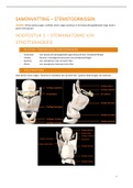
VON HERRICK
Doel: Diepte voorste oogkamer beoordelen
Methode
Optic section op precies 60º
Schijn het licht waar de cornea begint, net na de limbus
Vergelijk Dac met Dc
o Anterior chamber depth (Dac)
o Cornea Depth (Dc)
,
,CONICAL BEAM
Doel: Helderheid voorste oogkamer (VOK) bepalen. Controleren op cellen of flare (wittige
waas).
Methode
Kamer helemaal donker!
Spleethoogte 2-3mm
o Kleinste of één na kleinste rondje
Vergroting: Laag en Hoog
o Begin met 10x of 16x
o Vervolgens kijken met 25x en 40x
Verlichting spleetlamp maximum
40-50º graden
,ANTERIOR UVEITIS
Anterior uveïtis is inflammation involving the anterior uveal tract – the iris and anterior part
(pars plicata) of the ciliary body. The most common form of uveïtis.
Acute anteior uveïtis (AAU) is the most common presentation.
o Usually Idiopathic and HLA-B27 related (50% of cases)
o Secondary to inflammation elsewhere in the eye
Chronic anterior uveïtis (CAU) is less common.
o Bilateral, associated with systemic disease
o Associated with certain auto-immunity diseases
Spondyloarthropathies (SpA) – disorders which primarily affect the
joints
Ankylosing Spondylitis – involves lower spine problems
Psoriasis
Inflammatory bowel disease
Reactive athritis
o Associated with inflammatory diseases
Sarcoidosis – multisystem inflammatory disease
Idiopathic Juvenile Athritis – affecting children
Tubulointerstitial Nephritis – inflammatory kidney disorder
o Can be asymptomatic until the development of complications
Clinical Features (AAU)
Symptoms.
o Unilateral pain, photophobia, redness and watery discharge, miosis, headache
(one-sided)
o Commonly recurrent (history of similar episodes)
Visual Acuity.
o Only midly reduced.
o If severe, blurry vision
Ciliary Injection
o Circumcorneal conjunctival hyperaemia
o Purplish hue (involvement deeper blood vessels)
Anterior Chamber Cells
o Conical Beam
Estimate number of inflammatory cells in a 1mm by 1mm slit beam field
, Hypopyon
o Whitish purulent exudate composed of inflammatory cells in the inferior part of
the anterior chamber
o Common in HLA-B27-associated AAU
Keratic Precipitates (KP)
o Greyish-brown deposits on the corneal endothelium composed of inflammatory
cells
o Usually concentrated inferiorly
Aqueous Flare
o Haziness of the normally clear fluid in the anterior chamber (protein)
o Higher risk of complications
Intraocular Pressure (IOP)
o May be reduced at first (impairment aqeous secretion by cilairy epithelium)
o May be elevated later (inflammatory cells, etc)
Fibrinous Exudate
Iris Nodules
Posterior Syncechia (PS)
o Inflammatory adhesions between the pupil margin and the anterior lens capsule
o Pupil foten irregular and/or fixed
Iris Atrophy
Heterochromia Iridis
o Difference in colour between the iris of the two eyes
Iris Neovascularization (Rubeosis Iridis)
o Particularly in chronic inflammation
Cataract
o Mogelijke bijwerking door corticosteroïde gebruik
Investigation
Anamnese.
o Ask about any auto-immune disease – especially joint or lower back pain and
any skin rashes.
Spleetlamp
o Conical Beam
Helderheid VOK bepalen
Cellen en flare opsporen
o Oogdruk Meten
o Visus Meten
Treatment
Mydriatics or Cycloplegics
o Atropine or Cyclopentolate
1 or 2 drops
2 or 3 times a day
o To reduce pain and prevent complications
Prevents iris and ciliary muscle movement, reducing pain
Topical Corticosteroids
o Prednisolone Acetate 1% (Predforte) or Dexamethosone 0,1% (Maxidex)
Every 1-2 hrs, until inflammation is under control
Tapered over the next 4-6 wks
Check up weekly
o To control inflammation
Optometrie Jaar 2 Blok A
Elise de Ruiter
,Table of Contents
VOORSTE OOGKAMER ....................................................................................................................... 3
GEZICHTSBANEN EN DE NERVUS OPTICUS ................................................................................ 9
GLAUCOOM I – Sander Beers ........................................................................................................ 18
GLAUCOOM II – Sander Beers ....................................................................................................... 29
GLAUCOOM DEEL 1 .......................................................................................................................... 39
GLAUCOOM DEEL 2 .......................................................................................................................... 46
NEURO OPHTHALMOLOGIE .......................................................................................................... 56
ORBITA ................................................................................................................................................. 65
TONOMETRIE ..................................................................................................................................... 77
GONIOSCOPIE ..................................................................................................................................... 80
WERKCOLLEGES ................................................................................................................................ 84
ONDERZOEKSPLAN...................................................................................................................................... 84
GONIOSCOPIE ................................................................................................................................................ 84
EVALUATIE EN PLAN ................................................................................................................................... 85
,VOORSTE OOGKAMER
STUDIEDOELEN
Kanski – Uveïtis ‘Clinical Features’
Artikel – Optometric Grading Scales ‘Limbal Anterior Chamber Depth’
Artikel – Optometric Management of Anterior Segment Eye Disease
Vocabulair: KP, C/F, Synechia, Hypopyon
de diepte van de voorste oogkamer beoordelen met behulp van de van Herick methode.
de relatie van een nauwe en open kamerhoek met de van Herick methode uitleggen.
de risico’s weergeven bij de gradering van de van Herick methode.
de meting zelfstandig uitvoeren en noteren.
de oorzaken die een nauwe kamerhoek veroorzaken of versterken omschrijven.
het ziektebeeld uveïtis beoordelen en relateren aan klachten van de patiënt.
de relatie tussen de oogdruk en uveïtis omschrijven.
de relatie tussen systemische afwijkingen en uveïtis uitleggen.
ANATOMIE
(The lijn van schwalbe is het einde van Descemet’s membraan)
BEOORDELING DIEPTE VOORSTE OOGKAMER
Waarom?
Bepalen of het toedienen van pupilverwijdende druppels
mogelijk is.
Risico van kamerhoekafsluiting
o Acuut gesloten kamerhoekglaucoom.
Methodes
Penlight – minste nauwkeurig
Spleetlamp – Van Herrick
Gonisocopie – zich op kamerhoekstructuur
,PENLIGHT TEST
Hold the penlight parallel to the plane of the iris and temporally in order to direct the light
nasally.
Entire iris illuminated – anterior chamber is wide open (Grade 4)
Shadow – shallowness of anterior chamber (Grade 1 to 3)
VON HERRICK
Doel: Diepte voorste oogkamer beoordelen
Methode
Optic section op precies 60º
Schijn het licht waar de cornea begint, net na de limbus
Vergelijk Dac met Dc
o Anterior chamber depth (Dac)
o Cornea Depth (Dc)
,
,CONICAL BEAM
Doel: Helderheid voorste oogkamer (VOK) bepalen. Controleren op cellen of flare (wittige
waas).
Methode
Kamer helemaal donker!
Spleethoogte 2-3mm
o Kleinste of één na kleinste rondje
Vergroting: Laag en Hoog
o Begin met 10x of 16x
o Vervolgens kijken met 25x en 40x
Verlichting spleetlamp maximum
40-50º graden
,ANTERIOR UVEITIS
Anterior uveïtis is inflammation involving the anterior uveal tract – the iris and anterior part
(pars plicata) of the ciliary body. The most common form of uveïtis.
Acute anteior uveïtis (AAU) is the most common presentation.
o Usually Idiopathic and HLA-B27 related (50% of cases)
o Secondary to inflammation elsewhere in the eye
Chronic anterior uveïtis (CAU) is less common.
o Bilateral, associated with systemic disease
o Associated with certain auto-immunity diseases
Spondyloarthropathies (SpA) – disorders which primarily affect the
joints
Ankylosing Spondylitis – involves lower spine problems
Psoriasis
Inflammatory bowel disease
Reactive athritis
o Associated with inflammatory diseases
Sarcoidosis – multisystem inflammatory disease
Idiopathic Juvenile Athritis – affecting children
Tubulointerstitial Nephritis – inflammatory kidney disorder
o Can be asymptomatic until the development of complications
Clinical Features (AAU)
Symptoms.
o Unilateral pain, photophobia, redness and watery discharge, miosis, headache
(one-sided)
o Commonly recurrent (history of similar episodes)
Visual Acuity.
o Only midly reduced.
o If severe, blurry vision
Ciliary Injection
o Circumcorneal conjunctival hyperaemia
o Purplish hue (involvement deeper blood vessels)
Anterior Chamber Cells
o Conical Beam
Estimate number of inflammatory cells in a 1mm by 1mm slit beam field
, Hypopyon
o Whitish purulent exudate composed of inflammatory cells in the inferior part of
the anterior chamber
o Common in HLA-B27-associated AAU
Keratic Precipitates (KP)
o Greyish-brown deposits on the corneal endothelium composed of inflammatory
cells
o Usually concentrated inferiorly
Aqueous Flare
o Haziness of the normally clear fluid in the anterior chamber (protein)
o Higher risk of complications
Intraocular Pressure (IOP)
o May be reduced at first (impairment aqeous secretion by cilairy epithelium)
o May be elevated later (inflammatory cells, etc)
Fibrinous Exudate
Iris Nodules
Posterior Syncechia (PS)
o Inflammatory adhesions between the pupil margin and the anterior lens capsule
o Pupil foten irregular and/or fixed
Iris Atrophy
Heterochromia Iridis
o Difference in colour between the iris of the two eyes
Iris Neovascularization (Rubeosis Iridis)
o Particularly in chronic inflammation
Cataract
o Mogelijke bijwerking door corticosteroïde gebruik
Investigation
Anamnese.
o Ask about any auto-immune disease – especially joint or lower back pain and
any skin rashes.
Spleetlamp
o Conical Beam
Helderheid VOK bepalen
Cellen en flare opsporen
o Oogdruk Meten
o Visus Meten
Treatment
Mydriatics or Cycloplegics
o Atropine or Cyclopentolate
1 or 2 drops
2 or 3 times a day
o To reduce pain and prevent complications
Prevents iris and ciliary muscle movement, reducing pain
Topical Corticosteroids
o Prednisolone Acetate 1% (Predforte) or Dexamethosone 0,1% (Maxidex)
Every 1-2 hrs, until inflammation is under control
Tapered over the next 4-6 wks
Check up weekly
o To control inflammation