Case 1: Intra- and intercellular communication
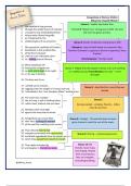
1. Explain types of signaling (auto-, para-, endocrine signalings, junction signalings,
juxtacrine signaling)
a. Categorize Delta-Notch, Wnt and Shh signaling pathways into the categories
above
b. cell to cell contact, paracrine - close range , endocrine through blood -long
range, autocrine - self, neighbouring
Juxtracrine signalling → ligand of one cell surface binds the other
Paracrine signalling → secreted molecules are carried to and act then on distant target cells
or act as local mediators. They can only act on cells in local environment of signalling cell
- signaling molecules rapidly taken up by neighbours
- signaling molecules destroyed by extracellular enzymes
- signaling molecules are immobilized in extracellular matrix
Endocrine signalling → long distance, molecules released from one cell, travels through
bloodstream to bind to distant target cell. Examples are hormones
Autocrine signalling → cell secretes hormone or chemical that binds the autocrine receptor
on the same cell, what leads to changes within the same cell
Intracrine signalling → hormone acts inside the cell and doesn’t leave the cell
Synaptic signaling → long distance, also paracrine since the signal is passed from one to
other cell. However, since neurons have long axons, this enables them to contact target cells
over long distances.
Notch signaling: autocrine or juxtracrine signaling
Wnt, Shh signaling: paracrine
2. How does cell to cell contact signalling work?
a. Look at: adherens junctions, tight junctions, gap junctions, delta-notch, desmosomes
and hemidesmosomes and actin linked desmosomes, Different types of Cadherins,
claudins and occludins, tricellulins, connexins and innexins, hemichannels and
connexons
1
,Cell to cell connections are essential in the following situations
- Epithelial cells of the skin → responsible for reshaping, flexibility and as barrier
- Epithelial cells of the gut → has barrier function and for flexibility
- Muscle cells → movement and strength
- Cardiomyocytes → heart function
Anchoring junction → is cell to cell adhesion and cell to matrix adhesion. This connection
can transmit stress and is tethered to cytoskeletal filaments inside the cell
The composition of this junction is mainly transmembrane adhesion proteins and intracellular
adaptor proteins which connect the intracellular filaments with the adhesion proteins
3 types of junctions:
➔ Adherens → actin filament attachment sites. Junction between cells.
These actin filaments pin the cells together that make sure the cells could move without
breaking. Cadherins are present on the membrane and interact in the space between the
two cells that interact and keeps both membranes together. Within the cells, cadherins
are attached to cytoplasmic plaques that connect intermediate filaments.
Cadherins depend on Ca2+ and are connected with Hinge regions. Ca2+ binds near
hinge to prevent it from flexing.
Cadherins can be found on the membranes of both cells that communicate with each
other and they interact in the space between them, and holds the 2 membranes
together. Inside the cell, cadherins attach to a structure called cytoplasmic plaque →
this connects the intermediate filaments and helps to anchor the junction
Cadherins depend on calcium. In the absence of Ca2+, cadherin behaves as a loose
and weak rope. When Ca2+ is present the molecules turn into stiff hook that links the
cells.
A cadherin molecule consists of several molecules connected by a hinge region. This
is also where the Ca2+ binds. Therefore it becomes stiff. When Ca2+ is removed the
hinges can flex and the molecule becomes floppy.
2
, ➔ Desmosomes → intermediate filament attachment sites. Junction between cells.
They also contain actin that allows stretch without breaking which provides
mechanical strength. They connect sites for bundles in intermediate filaments.
➔ Hemidesmosomes → intermediate filament attachment sites. Junction between cell
and matrix. Attaches to integrins and the integrins anchors the cell to laminin in basal
lamina what results in mechanical stability. The integrins span the membrane what
attaches keratin intracellularly via 2 adaptor proteins: plectin and BP230. Laminin is
used extracellularly to connect it to the collagen.
Occluding/tight junctions → seal gaps between cells in epithelium. The cell sheet is
impermeable or only selectively. This prevents molecules from one side of the sheet to the
other side of the sheet.
➔ Claudins → proteins that holds cells tightly against each other and interact with
partner group on opposite cell membrane. Main functions are
o Determine epithelial polarity by sequencing apical and basal domain
o Prevent free passage of substances across epithelial
o Connected to signaling networks that regulate cell differentiation
➔ Tricellular junctions → seal epithelium at corners of 3 cells
3
, Channel-forming junction → create passageways linking cytoplasm to adjacent cells.
➔ Gap junctions → 1.5nm water filled channels between 2 cells that allow transport of
ions, water ed. It is made out of connexins that form 1 connexon. Mainly present in
cardiac muscle where it helps to relay electrical signals since the ions of the action
potential can pass these gap junctions.
o Main function = mediation of the passage of chemical and electrical signals
from one to the other cell.
Signal-relaying junction → allow signals to be relayed from cell to cell across plasma
membranes at sites of cell to cell contact
4