Chapter 16 – Cel 2 CELBIOLOGIE
General principles of cell signaling:
- The signaling cell produces a particular type of extracellular signal molecule
that is detected by the target cell.
- Target cells process receptors that recognize and respond specifically to the
signaling molecule.
- Receptor produces intracellular signaling molecules: beginning signal
transduction.
Signaling molecules can be; proteins, peptides, amino acids, nucleotides,
steroids, fatty acid derivatives or even dissolved gases.
1. Hormones: extracellular signal molecules that signal through the whole
body by secreting it into an animal’s bloodstream.
2. Paracrine signaling: diffuse locally through the extracellular fluid. They
act as local mediators.
Many of the signal molecules that regulate inflammation at the site of an
infection function in this way.
3. Autocrine signaling: cells respond to the local mediators they have
produced themselves.
4. Neuronal signaling: nerve cells can deliver messages over long
distances. A message is targeted on target cells. They release a pulse of
an extracellular signal molecule: neurotransmitter.
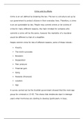
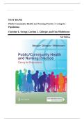
5. Signal-mediated cell-cell communication: most intimate and short
range, does not require the release of a secreted molecule. Cells make
direct physical contact through
signaling molecules lodged in the
plasma membrane of the target cell.
2 types of cell response: fast & slow:
Intracellular signaling pathways functions:
- They can relay the signal onward and
thereby help spread it through the cell.
- They can amplify the signal received,
making it stronger, so that a few
extracellular signal molecules are
enough to evoke a large intracellular
response.
- They can detect signals from more than one intracellular
signaling pathway and integrate them before relaying a
signal onward.
- They can distribute the signal to more than one effector
protein, creating branches in the information flow
diagram and evoking a complex response.
- They can modulate the response to the signal by
regulating the activity of components upstream in the
signaling pathway, a process known as feedback.
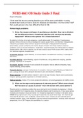
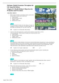
Molecular switches: receipt of a signal causes them to
toggle from an inactive to an active state. Activated; they can
stimulate/sometimes suppress other proteins in the signaling
pathway.
,For every step along the pathway there exists an inactivation mechanism.
Proteins that act as molecular switches fall mostly into one/two classes:
- Protein kinase: which covalently attaches a phosphate group onto the
switch protein.
2 main types:
- Serine/threonine kinases: phosphorylate proteins on serines or threonines.
- Tyrosine kinases: phosphorylate kinases.
- Protein phosphatase; which takes the phosphate off again.
These are often organized into phosphorylation cascades.
GTP-binding proteins: involved in intracellular signaling pathways. 2 main
types:
- Trimeric GTP-binding proteins (G-proteins): relay messages from G-
protein-coupled receptors.
- Monomeric GTPase: switch proteins are generally aided by 2 sets of
regulatory proteins that help them bind and hydrolyze GTP:
- Guanine nucleotide exchange factors (GEFs): activate switches by
promoting exchange GDP to GTP.
- GTPase-activating proteins (GAPs): turn them off by promoting GTP
hydrolysis.
Cell surface receptors, 3 main classes:
1. Ion-channel-coupled receptors: change the
permeability of the plasma membrane to selected
ions, thereby altering the membrane potential and
(under right conditions) producing an electrical
current.
2. G-protein-coupled receptors (GPCRs):
activate membrane-bound trimeric GTP-
binding proteins (G-proteins), which then
activate, or inhibit an enzyme or an ion
channel in the plasma membrane initiating
an intracellular signaling cascade.
When an extracellular signal molecule binds GPCRs; receptor undergoes
conformational change enables it to activate G protein.
G proteins
Some G proteins directly regulate ion channels: heartbeat; G protein activated
opens the K+ channel and inactivated keeps it closed.
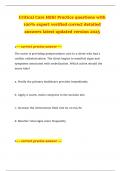
Interaction G proteins & enzymes: lead to production of additional
intracellular signaling molecules. The 2 most target enzymes of G
proteins:
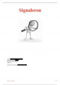
Adenylyl cyclase; produces small molecule called cyclin
AMP.
Cyclin AMP; the activated G protein α subunit switches on the
adenylyl cyclase, causing the increase synthesis of cyclic AMP from
ATP.
Exerts most of its effect by activating the enzyme cyclic-AMP-
dependent protein kinase (PKA). Active PKA catalyzes the
phosphorylation of particular serines or threonines on specific
intracellular proteins; altering its activity.
, The lower the cAMP levels are, the larger and faster the increase achieved
upon activation of adenylyl cyclase; which makes new cyclic AMP.
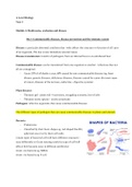
Phospholipase C; generates small molecules called inositol triphosphate
and diacylglycerol.
are activated by different G proteins; allowing production of small
molecules to different extracellular signals. These small molecules are
called: second messengers.
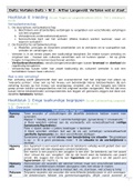
Activated phospholipase C propagates a signal by cleaving a lipid molecule that
is a component of the plasma membrane: inositol phospholipid. The cleavage
of the membrane inositol phospholipid generates 2 second messengers
molecules: (both play a crucial part in relaying the signal)
Inositol 1,4,5-triphosphate (IP3): a
water-soluble phosphate, is released
into cytosol where it binds Ca2+
channels in the ER. Ca2+ goes through
these channels causing a sharp rise in
the cytosol free Ca2+ concentration.
This gives signals to other proteins.
Diacylglycerol (DAG): lipid that
remains in the plasma membrane. It
helps recruit and activate protein
kinase C (PKC). PKC translocases from
the cytosol to the plasma membrane. It
needs to bind Ca2+ to become active.
Activated PKC phosphorylates a set of
intracellular proteins.
If Ca2+ channels open, Ca2+ rushes down the electrochemical gradient
into the cytosol, where it triggers changes in Ca2+-responsive proteins.
Effect of Ca2+ on Ca2+-responsive proteins in cytosol are largely indirect:
Calmodulin: when Ca2+ binds to calmodulin; the protein undergoes a
conformational change that enables it to interact with target proteins in
the cell altering their activities. One important class of targets is the
Ca2+/calmodulin-dependent protein kinases (CaM-kinases). When
activated, they influence other processes by phosphorylating selected
proteins. The CaM-kinase is activated by the pulses of Ca 2+ signals that
occur during neural activity.
GPCR generates a dissolved gas that carries a signal to adjacent cells; no second
messenger needed.
Example: nitric oxide (NO): acts as a signaling molecule in many tissues. It
diffuses readily from its site of synthesis and slips into neighboring cells. The
distance is limited; bc of the interaction oxygen and water that makes NO
nitrates and nitrites.
Effect NO on blood vessels: Ca2+ stimulates the NO synthase, this NO diffuses into
smooth muscle cells in the vessel to dilate, so that blood flows through it more
freely. Nitroglycerin is converted to NO and causes the relax of the blood vessels.
NO is also used to signal neighboring cells. Inside target cells it binds and
activates the enzyme guanylyl cyclase, stimulating the formation of cyclic GMP
(second messenger) from GTP.
Adaption: occurs in intracellular signaling pathways that respond to extracellular
signal molecules, allowing cells to respond to fluctuations in the concentration of
such molecules regardless of whether they are present in small or large amounts.
General principles of cell signaling:
- The signaling cell produces a particular type of extracellular signal molecule
that is detected by the target cell.
- Target cells process receptors that recognize and respond specifically to the
signaling molecule.
- Receptor produces intracellular signaling molecules: beginning signal
transduction.
Signaling molecules can be; proteins, peptides, amino acids, nucleotides,
steroids, fatty acid derivatives or even dissolved gases.
1. Hormones: extracellular signal molecules that signal through the whole
body by secreting it into an animal’s bloodstream.
2. Paracrine signaling: diffuse locally through the extracellular fluid. They
act as local mediators.
Many of the signal molecules that regulate inflammation at the site of an
infection function in this way.
3. Autocrine signaling: cells respond to the local mediators they have
produced themselves.
4. Neuronal signaling: nerve cells can deliver messages over long
distances. A message is targeted on target cells. They release a pulse of
an extracellular signal molecule: neurotransmitter.
5. Signal-mediated cell-cell communication: most intimate and short
range, does not require the release of a secreted molecule. Cells make
direct physical contact through
signaling molecules lodged in the
plasma membrane of the target cell.
2 types of cell response: fast & slow:
Intracellular signaling pathways functions:
- They can relay the signal onward and
thereby help spread it through the cell.
- They can amplify the signal received,
making it stronger, so that a few
extracellular signal molecules are
enough to evoke a large intracellular
response.
- They can detect signals from more than one intracellular
signaling pathway and integrate them before relaying a
signal onward.
- They can distribute the signal to more than one effector
protein, creating branches in the information flow
diagram and evoking a complex response.
- They can modulate the response to the signal by
regulating the activity of components upstream in the
signaling pathway, a process known as feedback.
Molecular switches: receipt of a signal causes them to
toggle from an inactive to an active state. Activated; they can
stimulate/sometimes suppress other proteins in the signaling
pathway.
,For every step along the pathway there exists an inactivation mechanism.
Proteins that act as molecular switches fall mostly into one/two classes:
- Protein kinase: which covalently attaches a phosphate group onto the
switch protein.
2 main types:
- Serine/threonine kinases: phosphorylate proteins on serines or threonines.
- Tyrosine kinases: phosphorylate kinases.
- Protein phosphatase; which takes the phosphate off again.
These are often organized into phosphorylation cascades.
GTP-binding proteins: involved in intracellular signaling pathways. 2 main
types:
- Trimeric GTP-binding proteins (G-proteins): relay messages from G-
protein-coupled receptors.
- Monomeric GTPase: switch proteins are generally aided by 2 sets of
regulatory proteins that help them bind and hydrolyze GTP:
- Guanine nucleotide exchange factors (GEFs): activate switches by
promoting exchange GDP to GTP.
- GTPase-activating proteins (GAPs): turn them off by promoting GTP
hydrolysis.
Cell surface receptors, 3 main classes:
1. Ion-channel-coupled receptors: change the
permeability of the plasma membrane to selected
ions, thereby altering the membrane potential and
(under right conditions) producing an electrical
current.
2. G-protein-coupled receptors (GPCRs):
activate membrane-bound trimeric GTP-
binding proteins (G-proteins), which then
activate, or inhibit an enzyme or an ion
channel in the plasma membrane initiating
an intracellular signaling cascade.
When an extracellular signal molecule binds GPCRs; receptor undergoes
conformational change enables it to activate G protein.
G proteins
Some G proteins directly regulate ion channels: heartbeat; G protein activated
opens the K+ channel and inactivated keeps it closed.
Interaction G proteins & enzymes: lead to production of additional
intracellular signaling molecules. The 2 most target enzymes of G
proteins:
Adenylyl cyclase; produces small molecule called cyclin
AMP.
Cyclin AMP; the activated G protein α subunit switches on the
adenylyl cyclase, causing the increase synthesis of cyclic AMP from
ATP.
Exerts most of its effect by activating the enzyme cyclic-AMP-
dependent protein kinase (PKA). Active PKA catalyzes the
phosphorylation of particular serines or threonines on specific
intracellular proteins; altering its activity.
, The lower the cAMP levels are, the larger and faster the increase achieved
upon activation of adenylyl cyclase; which makes new cyclic AMP.
Phospholipase C; generates small molecules called inositol triphosphate
and diacylglycerol.
are activated by different G proteins; allowing production of small
molecules to different extracellular signals. These small molecules are
called: second messengers.
Activated phospholipase C propagates a signal by cleaving a lipid molecule that
is a component of the plasma membrane: inositol phospholipid. The cleavage
of the membrane inositol phospholipid generates 2 second messengers
molecules: (both play a crucial part in relaying the signal)
Inositol 1,4,5-triphosphate (IP3): a
water-soluble phosphate, is released
into cytosol where it binds Ca2+
channels in the ER. Ca2+ goes through
these channels causing a sharp rise in
the cytosol free Ca2+ concentration.
This gives signals to other proteins.
Diacylglycerol (DAG): lipid that
remains in the plasma membrane. It
helps recruit and activate protein
kinase C (PKC). PKC translocases from
the cytosol to the plasma membrane. It
needs to bind Ca2+ to become active.
Activated PKC phosphorylates a set of
intracellular proteins.
If Ca2+ channels open, Ca2+ rushes down the electrochemical gradient
into the cytosol, where it triggers changes in Ca2+-responsive proteins.
Effect of Ca2+ on Ca2+-responsive proteins in cytosol are largely indirect:
Calmodulin: when Ca2+ binds to calmodulin; the protein undergoes a
conformational change that enables it to interact with target proteins in
the cell altering their activities. One important class of targets is the
Ca2+/calmodulin-dependent protein kinases (CaM-kinases). When
activated, they influence other processes by phosphorylating selected
proteins. The CaM-kinase is activated by the pulses of Ca 2+ signals that
occur during neural activity.
GPCR generates a dissolved gas that carries a signal to adjacent cells; no second
messenger needed.
Example: nitric oxide (NO): acts as a signaling molecule in many tissues. It
diffuses readily from its site of synthesis and slips into neighboring cells. The
distance is limited; bc of the interaction oxygen and water that makes NO
nitrates and nitrites.
Effect NO on blood vessels: Ca2+ stimulates the NO synthase, this NO diffuses into
smooth muscle cells in the vessel to dilate, so that blood flows through it more
freely. Nitroglycerin is converted to NO and causes the relax of the blood vessels.
NO is also used to signal neighboring cells. Inside target cells it binds and
activates the enzyme guanylyl cyclase, stimulating the formation of cyclic GMP
(second messenger) from GTP.
Adaption: occurs in intracellular signaling pathways that respond to extracellular
signal molecules, allowing cells to respond to fluctuations in the concentration of
such molecules regardless of whether they are present in small or large amounts.