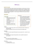
fMRI Scans
How DO they work?
The head is placed inside a large and powerful electromagnet. As neural activity in the brain
increases so does the blood flow to keep up with the oxygen demand. The oxygen is carried
to the neurons by haemoglobin within red blood cells. When it carries oxygen haemoglobin
repeals a magnetic field (diamagnetic) but when it’s deoxygenated it follows the direction of
the magnetic field (paramagnetic). It is these changes in direction that the scanner detects
to create an image.
Evaluation:
Weaknesses:
Strengths: o The use of high-powered magnetic
o Enables images of brain activity fields mean that some people are
to be gathered without the use unable to have these kind of brain
of radiation. scans, for example: someone with a
o Comparative safety to other cardiac pacemaker or who has had a
forms of scans. metal surgical implant.
o The procedure of choice for o The procedure may not be suitable
psychologists. for someone who is claustrophobic
o Non-invasive, so doesn’t share or is unnerved by confined spaces.
the potential risks of PET scans. This because they have to lie flat
and still in a large tube for the
duration of the scan.
Validity:
▪ Provides a detailed image of both structure and function however movement can
distort the image.
▪ Objective interpretation of bot structure and function (red=high activity)
Reliability:
▪ High- can be easily repeated following the same procedure (time + number of scans)
Generalisability:
▪ Cannot be used for people with pacemakers and not suitable for people with
claustrophobia.
Ethics:
▪ Non-invasive and no radiation exposure.
▪ Issues with claustrophobia and scanning method is loud.
Time/Cost:
▪ Costs around £500
▪ Time efficient (15-60 minutes)
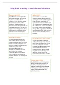