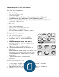
Zebrafish genetics and development
Zebrafish as a model organism
- Size: 3 cm long
- Diet: brine shrimp and algae
- Life span: ~5 years
- Genome size: 1700 Mb (25 diploid – unlike many other fish, ~24000 genes)
- Vertebrate model, rapid development, optically transparent embryo
- Forward, reverse and transgenic approaches, easily injected, easy to make mosaics
- Conservation of gene function
Used to study:
- Visible internal development
- Cardiovascular and neurological studies
- Eye development and eye diseases
- Gene knockdown (morpholino injection in embryo)
Stages of zebrafish development
Cells divide on top of yolk
Cleavage divisions followed by epiboly (~6h)
Involution to form the internal endoderm and
mesoderm
Convergent-extension: forms body axis and
extending the embryo, cells flow in one direction
causing extension
- Migration of anterior paraxial mesoderm
Technological advances allow better imaging and
observation
- SPIM (light-sheet imaging): allows quick imaging in low
toxicity environment, super-resolution, high-speed
- Fluorescent transgene to allow tracking of nuclei –
extract data
- Cell division and imaging – can show tissue
development
Identifying developmental genes
Forward Genetic Screens – identify mutant phenotypes via F1/F2 screens
(phenotype then work back to identify gene)
Reverse genetic screens – knock out the gene, observe effect on phenotype
Saturation screens – isolation of more than 4000 mutations
, Some phenotypes include: cyclopia/holoproencephaly (morphological), subtle
phenotypes (look at connectivity in brain nerves), Paz2 mutation leads to defects in
optic axon guidance (mutations in orthologous genes of mice and humans lead to
similar defects, behavioural
Mutations leading to defects in commissure formation in the forebrain
Many single gene mutations don’t give strong phenotypes…
- Forward genetic screens now look for
interactions between genes
- Fish already carrying mutations that
are susceptible to certain phenotypes –
continue breeding
- Then observe the effect of the loss of
2 genes (eg. c is a double mutant with
much smaller eyes)
- Easy to do as females lay many eggs
An issue: FGS have identified ~10,000
mutant but causal genes is hard to
identify (~9900 mutants, but the gene is only known for ~30% of them)
Identifying the Causal Gene
a) Mapping and positional cloning
Linkage mapping – utilises the fact that genes that are
further away are more likely to produce recombinant
phenotypes due to crossover during meiosis
Determine how frequently recombination occurs between
markers during meiosis
What markers do use? SNPs, SSLPs (aka CA-repeats, SSRs,
microsatellites)
Length of CA tract differs between individuals
- Co-dominant: good for haploid and diploid crosses
- Informative in most crosses: 50-90%
- Robust markers: easily scored by PCR, reproducible banding patterns, easy to
transfer information between crosses and labs
b) Transcriptome or genome sequencing based identification of mutations (as
zebrafish genome has been fully sequenced)
- Next-Generation Sequencing to Map Mutations – homozygosity mapping
- Basic principle the same – if recombinants rare, the SNP is close to the mutant
allele
Testing candidate genes
Anti-sense approach: Morpholinos block translation (can also block splicing)
, Newer approaches – finding mutations in identified genes
- Traditional Use a chemical mutagen to induce mutations in the genome
(cryopreserve sperm from F1 mutan ts, then sequence the DNA)
- After identifying mutatations using assay – recover mutant line from frozen sperm
(selecting fish with particular mutations)
New approach – hijack DNA repair mechnanism
Homology directed repair – find sister chromatid, use that to repair damaged DNA
(longer process, but more precise)
NHEJ - Non-homologous end-joining
Zinc finger nucleases – zinc finger preferentially binds to certain DNA sequences .
can obtain cut at a specific site (needed to test many different zinc finger
sequences to get the correct one)
Talens: Isolate repetitive sequence and a variable section
CRISPRs: specificity provided by RNA, guide RNA (complementary to target DNA),
recognise the target DNA, cas9 binds to guide RNA which then cuts the DNA.
Imprecise repair – can introduce indels targeted genome editing
Applications of CRISPRs –indel, large insertions or replacement, large deletions or
rearrangement, couple cas9 with transcriptional activation domain – can activate
genes
Chimaeras – cells moved around (take cells from one embryo, labelled, then
transplant to another embryo)
Applications – small molecule screening in zebrafish embryos
Add a molecule in plate – observed the phenotype (pigment is removed, used to
study melanoma etc)
Drug discovery – Use 700 types of drugs and treat embryos, observe effect on
melanocytes
Inflammation
Dynamics of process can be studied – zebrafish transparent (no need for transgene
or cell labelling)
Blood cell populations can be labelled with GFP (eg. macrophages) can see how cells
are recruited to the wound site
Brain development
Different parts of body have different levels of sensitivity – can observed the
density of nerves in the embryo
Sensory neuromasts, lateral line primordium (study collective cell movement)
Multiple labels allow the types of cells to be distinguished