1. Skeletal system
Functions:
Production of blood
Fat storage
Transmit sound
Gives body its basic shape, supports allows body to stay up right
Protects basic and vital organs
Pivot or rotate according to muscle movement
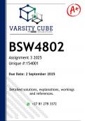
Structure:
Bone is living tissue
Compacts bone – hard/dense tissue used for weight bearing abilities. It forms the surface/
midsection of bones (long bone). Layers (periosteum membrane that covers the outer bone
and provides nutrients and o2. Where tendons attach
Spongey bone – cancellous bone. Shorter/lighter bone tissue. Heads of long bones. Contains
red bone marrow (red blood cells, white blood cells production). Exerts resistance against
forces that act on the bone (shock absorber)
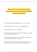
Type of bones
Long bone – movement, support, red blood cell production (femur/ humorous/ tibia/ radius/
ulna)
Shot bone – fine or small movement, stability, weight bearing, shock absorbent (carpals/
tarsals)
Flat bones – attachment for muscles and for production (sternum/scapular/ pelvis/ cranium)
Sesamoid – protection, reduction of friction across a joint (patella/ wrist)
Irregular bones – protection and movement (vertebra)
Ossification:
Bone formation (hardening of bone as you develop)
Begins at 3 weeks old, bone starts off as cartilage, osteoblasts invade cartilage and produce
osteoid (collagen with sites for calcium phosphate crystals to form, giving the bone strength)
Process begins in middle of bone (primary ossification center), process continues outward.
Blood vessels then grow into bones reshaped by osteoclasts so there is a bone marrow filled
central cavity.
Growth plates at the end of each long bone allowing more bone to be produced in order to
grow through child hood. Growth plate lose and become bone after growth is completed.
Growth Hormones- sent from pituitary gland to start process. Stimulates cartilage
production.
Cartilage – semitransparent tough and elastic connective tissue
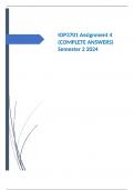
Joints:
2 or more bones meet (articulation). have shock absorbers made of bags of squishy fluid.
Fixed/ fibrous joints - stable and no visible movement, lots of connective tissue (skull)
Cartilaginous joints – slightly movable. Joint by tough cartilage providing stability and shock
absorption (vertebra disks and pelvic bone)
, Synovial joints- freely moveable, covered in cartilage, sharp absorbent ligaments
1. Gliding joint – 2 flat surfaces glide past each other, are biaxial and allow movement in all
directions. (wrist/ carpal bones)
2. ellipsoid – 2 plane movement back and forth
3. hinge joint – uniaxial, 1 plane movement ligaments prevent other movement (knee)
4. pivot joint – uniaxial but allow rotation (cervical vertebra)
5. saddle joint – biaxial where concave and convex surfaces meet (thumb)
6. ball and socket joint – biggest range of movement, round bone into cup shaped cavity
(hip/shoulder)
movement – flexion, extension, dorsiflexion, adduction and abduction. Bend – flexors
straighten – extensors.
Connective tissue:
Ligaments – tough fibrous connective tissue, attaches one bone to another, stretches
enough to control movement of joint and stabilizes it. Not elastic.
Tendons – attach muscle to bone, strong and inelastic, small – don’t have nerves, big –
connected to CNS and CS for nerves and blood supply.
Bone issues:
Aging – less synovial fluid produced causing brittle bones and stiffness
Poor dietary habits
Deficiency of nutrients and minerals
Prior accidents increase risk of fractures
2. Muscular system
, Structure:
Tissue – smooth muscle tissue and cardiac muscle are involuntary (actin
continuously without thought and doesn’t tire)
Skeletal muscle tissue – voluntary control (actin done continuously)
Skeletal muscle
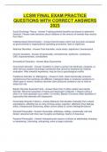
Muscle filament structure:
Made of lots of tiny fibers in bundles. Muscles contract and relax against each other
Fiber is surrounded by membrane called sarcolemma. Inside cells are myofibrils
(long, tubular structure running the length of muscle). They are imbedded in cells
sarcoplasm.
Arranged in a regular stripped pattern.
Sarcomere (makes muscles contract) = myofibrils separated into units called
contractile units.
Thick filament – protein myosin (myosin heads)
Thin filament – protein actin (binding sites)
Muscle bundle Held together by collagen and combined by connective tissue
(epimysium)
Muscle spindles (intrafusal muscle fibers)
Sensory nerves in body of muscle. Direct change in muscle length activates motor
neurons. 2 sensory endings; 1: primary (respond to speed and size of muscle length
change. 2: secondary (respond to amount of stretch)
How movement happens:
Nerves meet muscle at neuromuscular junction. Electrical signal causes myofilaments to
slide across each other, sarcomere then shortens to generate force
Actin and myosin provide all muscle movement when slide together (contractions) and apart
(relaxation)
Sensory nerves in body of muscle need to be stimulated by CNS. Direct change in muscle
length activates motor neurons. 2 sensory endings; 1: primary (respond to speed and size of
muscle length change. 2: secondary (respond to amount of stretch)
Motor unit – stimulate group of muscle fibers motor unit is made up of single neuron in
order to activate, signal from spinal cord reaches motor unit and enables it to contract.
Muscle spindles: (will be continued in grd 12)
Detect changes in length of muscle and assist in regulating contractions by activation motor
neurons.
Structure – small sensory organs, elongated, consist of several modified muscle fibers
enclosed in connective tissue
2 sensory endings; 1: primary (respond to speed and size of muscle length change. 2:
secondary (respond to amount of stretch). Endings located in the middle of spindle
Functions:
Production of blood
Fat storage
Transmit sound
Gives body its basic shape, supports allows body to stay up right
Protects basic and vital organs
Pivot or rotate according to muscle movement
Structure:
Bone is living tissue
Compacts bone – hard/dense tissue used for weight bearing abilities. It forms the surface/
midsection of bones (long bone). Layers (periosteum membrane that covers the outer bone
and provides nutrients and o2. Where tendons attach
Spongey bone – cancellous bone. Shorter/lighter bone tissue. Heads of long bones. Contains
red bone marrow (red blood cells, white blood cells production). Exerts resistance against
forces that act on the bone (shock absorber)
Type of bones
Long bone – movement, support, red blood cell production (femur/ humorous/ tibia/ radius/
ulna)
Shot bone – fine or small movement, stability, weight bearing, shock absorbent (carpals/
tarsals)
Flat bones – attachment for muscles and for production (sternum/scapular/ pelvis/ cranium)
Sesamoid – protection, reduction of friction across a joint (patella/ wrist)
Irregular bones – protection and movement (vertebra)
Ossification:
Bone formation (hardening of bone as you develop)
Begins at 3 weeks old, bone starts off as cartilage, osteoblasts invade cartilage and produce
osteoid (collagen with sites for calcium phosphate crystals to form, giving the bone strength)
Process begins in middle of bone (primary ossification center), process continues outward.
Blood vessels then grow into bones reshaped by osteoclasts so there is a bone marrow filled
central cavity.
Growth plates at the end of each long bone allowing more bone to be produced in order to
grow through child hood. Growth plate lose and become bone after growth is completed.
Growth Hormones- sent from pituitary gland to start process. Stimulates cartilage
production.
Cartilage – semitransparent tough and elastic connective tissue
Joints:
2 or more bones meet (articulation). have shock absorbers made of bags of squishy fluid.
Fixed/ fibrous joints - stable and no visible movement, lots of connective tissue (skull)
Cartilaginous joints – slightly movable. Joint by tough cartilage providing stability and shock
absorption (vertebra disks and pelvic bone)
, Synovial joints- freely moveable, covered in cartilage, sharp absorbent ligaments
1. Gliding joint – 2 flat surfaces glide past each other, are biaxial and allow movement in all
directions. (wrist/ carpal bones)
2. ellipsoid – 2 plane movement back and forth
3. hinge joint – uniaxial, 1 plane movement ligaments prevent other movement (knee)
4. pivot joint – uniaxial but allow rotation (cervical vertebra)
5. saddle joint – biaxial where concave and convex surfaces meet (thumb)
6. ball and socket joint – biggest range of movement, round bone into cup shaped cavity
(hip/shoulder)
movement – flexion, extension, dorsiflexion, adduction and abduction. Bend – flexors
straighten – extensors.
Connective tissue:
Ligaments – tough fibrous connective tissue, attaches one bone to another, stretches
enough to control movement of joint and stabilizes it. Not elastic.
Tendons – attach muscle to bone, strong and inelastic, small – don’t have nerves, big –
connected to CNS and CS for nerves and blood supply.
Bone issues:
Aging – less synovial fluid produced causing brittle bones and stiffness
Poor dietary habits
Deficiency of nutrients and minerals
Prior accidents increase risk of fractures
2. Muscular system
, Structure:
Tissue – smooth muscle tissue and cardiac muscle are involuntary (actin
continuously without thought and doesn’t tire)
Skeletal muscle tissue – voluntary control (actin done continuously)
Skeletal muscle
Muscle filament structure:
Made of lots of tiny fibers in bundles. Muscles contract and relax against each other
Fiber is surrounded by membrane called sarcolemma. Inside cells are myofibrils
(long, tubular structure running the length of muscle). They are imbedded in cells
sarcoplasm.
Arranged in a regular stripped pattern.
Sarcomere (makes muscles contract) = myofibrils separated into units called
contractile units.
Thick filament – protein myosin (myosin heads)
Thin filament – protein actin (binding sites)
Muscle bundle Held together by collagen and combined by connective tissue
(epimysium)
Muscle spindles (intrafusal muscle fibers)
Sensory nerves in body of muscle. Direct change in muscle length activates motor
neurons. 2 sensory endings; 1: primary (respond to speed and size of muscle length
change. 2: secondary (respond to amount of stretch)
How movement happens:
Nerves meet muscle at neuromuscular junction. Electrical signal causes myofilaments to
slide across each other, sarcomere then shortens to generate force
Actin and myosin provide all muscle movement when slide together (contractions) and apart
(relaxation)
Sensory nerves in body of muscle need to be stimulated by CNS. Direct change in muscle
length activates motor neurons. 2 sensory endings; 1: primary (respond to speed and size of
muscle length change. 2: secondary (respond to amount of stretch)
Motor unit – stimulate group of muscle fibers motor unit is made up of single neuron in
order to activate, signal from spinal cord reaches motor unit and enables it to contract.
Muscle spindles: (will be continued in grd 12)
Detect changes in length of muscle and assist in regulating contractions by activation motor
neurons.
Structure – small sensory organs, elongated, consist of several modified muscle fibers
enclosed in connective tissue
2 sensory endings; 1: primary (respond to speed and size of muscle length change. 2:
secondary (respond to amount of stretch). Endings located in the middle of spindle