Sunday, 4 September y
Cells
Cell structure
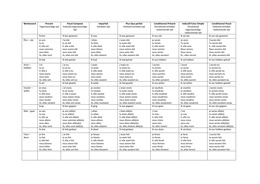
Eukaryote Prokaryote Virus
Cell membrane Cell membrane Capsid
Cell wall Envelope derived from host cell
Capsule membrane (removal of too much
from the host destroys the host
as it doesn't have enough mem-
brane for itself)
DNA in nucleus Loop of DNA and plasmids viral DNA/RNA
Phospholipids - cell membrane
Glycoproteins e.g murein - cell
wall
Cells under a microscope
Cells can look different under a microscope as…
- it is a cross section
- Organelles can be orientated differently
Light microscope cannot see
- Mitochondria
- RER
- Cell membrane
- SER
- lysosomes
- Ribosomes
Mitochondria
- site of aerobic respiration
- produces ATP
- matrix contains DNA (stores substrates for protein synthesis) (synthesises enzymes
for respiration)
Chloroplast function
1
, Sunday, 4 September y
- absorb light - site of light dependent reaction (contains chlorophyll - photosynthetic
pigments)(grana increase s/a to maximise light exposure)(starch granules act as food
storage)
- photosynthesis
- produce sugars (light independent reaction)
Lysosomes
- membrane bound sac - containing digestive enzymes
- secretes these to break down pathogens in phagocytes
Specialised cells
Xylem cell
- less cell content > free movement of water
- Walls between cells break down > continuous column of water
- Cell walls are strengthened with lignin > prevents damage to xylem vessel
Root hair cell
- large s/a >
more water
ab- sorption
- many mito-
chon- dria >
ATP for ac-
tive trans-
port of min-
erals
Cellu- lar or-
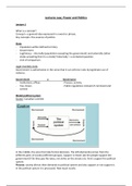
ganisa- tion
Cells > tissues
> or- gans >
organ systems
2
, Sunday, 4 September y
Structures in fig. 1 that are in eukaryotes but not prokaryotes
- nucleus
- Starch
- Eye spot
Fig 1 =
a producer - chloroplast for photosynthesis
Eukaryote - primitive sight organ
The microscope in cell studies
Optical microscope TEM SEM
Image formed using… Light Electrons Electrons
Resolution Low Highest High
Maximum magnification x1500 X1,500,000 X1,500,000
Used to observe Nucleus Nucleus Cell surface
Mitochondria
Lysosomes
RER
plasma membrane
SER
Ribosomes
Image 2D 2D 3D
Colour Colour B+W B+W
Sample thickness must be thin must be thin can be solid
Sample type can be alive must be dead (in a vac- Must be dead (in a vac-
uum) uum)
Theoretical resolving power of EMs not always reached..
3
Cells
Cell structure
Eukaryote Prokaryote Virus
Cell membrane Cell membrane Capsid
Cell wall Envelope derived from host cell
Capsule membrane (removal of too much
from the host destroys the host
as it doesn't have enough mem-
brane for itself)
DNA in nucleus Loop of DNA and plasmids viral DNA/RNA
Phospholipids - cell membrane
Glycoproteins e.g murein - cell
wall
Cells under a microscope
Cells can look different under a microscope as…
- it is a cross section
- Organelles can be orientated differently
Light microscope cannot see
- Mitochondria
- RER
- Cell membrane
- SER
- lysosomes
- Ribosomes
Mitochondria
- site of aerobic respiration
- produces ATP
- matrix contains DNA (stores substrates for protein synthesis) (synthesises enzymes
for respiration)
Chloroplast function
1
, Sunday, 4 September y
- absorb light - site of light dependent reaction (contains chlorophyll - photosynthetic
pigments)(grana increase s/a to maximise light exposure)(starch granules act as food
storage)
- photosynthesis
- produce sugars (light independent reaction)
Lysosomes
- membrane bound sac - containing digestive enzymes
- secretes these to break down pathogens in phagocytes
Specialised cells
Xylem cell
- less cell content > free movement of water
- Walls between cells break down > continuous column of water
- Cell walls are strengthened with lignin > prevents damage to xylem vessel
Root hair cell
- large s/a >
more water
ab- sorption
- many mito-
chon- dria >
ATP for ac-
tive trans-
port of min-
erals
Cellu- lar or-
ganisa- tion
Cells > tissues
> or- gans >
organ systems
2
, Sunday, 4 September y
Structures in fig. 1 that are in eukaryotes but not prokaryotes
- nucleus
- Starch
- Eye spot
Fig 1 =
a producer - chloroplast for photosynthesis
Eukaryote - primitive sight organ
The microscope in cell studies
Optical microscope TEM SEM
Image formed using… Light Electrons Electrons
Resolution Low Highest High
Maximum magnification x1500 X1,500,000 X1,500,000
Used to observe Nucleus Nucleus Cell surface
Mitochondria
Lysosomes
RER
plasma membrane
SER
Ribosomes
Image 2D 2D 3D
Colour Colour B+W B+W
Sample thickness must be thin must be thin can be solid
Sample type can be alive must be dead (in a vac- Must be dead (in a vac-
uum) uum)
Theoretical resolving power of EMs not always reached..
3