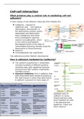
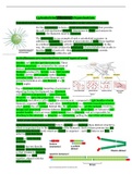
Cell-cell interaction
What proteins play a central role in mediating cell-cell
adhesion?
3 main classes of cell adhesion molecules that mediate this:
Cadherins – classical (E
cadherins) and non-classical
(desmoglein, desmocollin). In
the desmosome junction shown,
desmoglein and desmocollin
have cytoplasmic tails which
bind to plakoglobin (gamma-
catenin) and plakophilin, which
in turn bind to desmoplakin.
Desmoplakin binds to the sides of
intermediate filaments, thereby tying the
desmosome to these filaments.
Selectins
Immunoglobulin (Ig) superfamily
Top: adherens junction. Bottom: desmosome
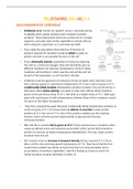
How is adhesion mediated by Cadherins?
The cadherin superfamily in vertebrates
includes hundreds of different proteins,
including many with signalling functions.
Humans have 180 cadherin superfamily
members/ genes.
Classical cadherins: first 3 cadherins that
were discovered were named according to
the main tissues in which they were found.
E-cadherin is present on many types of
epithelial cells; N-
cadherin on nerve,
muscle and lens cells;
and P-cadherin on cells
in the placenta and
epidermis. These and
other classical
, cadherins are closely related in sequence, and while they all have
well-defined adhesive functions, they are also important in
signalling.
Non-classical cadherins: more distantly related in sequence.
Include proteins with known adhesive function, such as
protocadherins found in the brain, and desmocollins & desmogleins
that form desmosome junctions. They also include proteins primarily
involved in signalling, such as T-cadherin in nerve and muscle cells
and the Fat and Flamingo (latter involved in signal transduction).
Large diversity among cadherin superfamily members: these
proteins all have extracellular portions containing multiple
copies of the cadherin domain motif, but their intracellular
portions are more varied, reflecting interactions with a wide
variety of intracellular ligands, including signalling molecules
as well as components that anchor the cadherin to the
cytoskeleton. The differently coloured motifs in Fat, Flamingo
and Ret represent conserved domains that are also found in
other protein families.
Cadherins associate through homophillic interactions:
Binding between cadherins is generally homophillic (like-to-
like): cadherin molecules of a specific subtype on one cell bind
to cadherin molecules of the same/ closely related subtype on
adjacent cells. E.g. E-cadherin with E-cadherin
This binding occurs at the N-terminal tips of cadherin molecules –
furthest from the membrane.
Some other cell adhesion molecules bind heterophilically (e.g.
integrins, selectins) – different molecules interact with each other.
Cadherin-mediated adhesions are calcium-dependent:
2 classical cadherin molecules on opposite
cells bind homophilically, end-to-end. This
explains why this interaction is relatively
weak (cadherins bind to their partners
with low affinity, strong attachments
forming from many weak bonds in
parallel). Means that dynamic adjustment
of adhesion can be made by controlling
cadherin expression level and trafficking to/from membrane.
The extracellular part of each polypeptide consists of a series of
compact domains called cadherin repeats, joined by a flexible hinge
region. Ca2+ binds in the region of each hinge,
preventing it from flexing. In the absence of Ca2+, the
molecule becomes floppy and adhesion fails.
Outside cells, calcium ion levels are high, > 1mM. If
they deplete to less than 0.05mM, Ca2+ leave the
What proteins play a central role in mediating cell-cell
adhesion?
3 main classes of cell adhesion molecules that mediate this:
Cadherins – classical (E
cadherins) and non-classical
(desmoglein, desmocollin). In
the desmosome junction shown,
desmoglein and desmocollin
have cytoplasmic tails which
bind to plakoglobin (gamma-
catenin) and plakophilin, which
in turn bind to desmoplakin.
Desmoplakin binds to the sides of
intermediate filaments, thereby tying the
desmosome to these filaments.
Selectins
Immunoglobulin (Ig) superfamily
Top: adherens junction. Bottom: desmosome
How is adhesion mediated by Cadherins?
The cadherin superfamily in vertebrates
includes hundreds of different proteins,
including many with signalling functions.
Humans have 180 cadherin superfamily
members/ genes.
Classical cadherins: first 3 cadherins that
were discovered were named according to
the main tissues in which they were found.
E-cadherin is present on many types of
epithelial cells; N-
cadherin on nerve,
muscle and lens cells;
and P-cadherin on cells
in the placenta and
epidermis. These and
other classical
, cadherins are closely related in sequence, and while they all have
well-defined adhesive functions, they are also important in
signalling.
Non-classical cadherins: more distantly related in sequence.
Include proteins with known adhesive function, such as
protocadherins found in the brain, and desmocollins & desmogleins
that form desmosome junctions. They also include proteins primarily
involved in signalling, such as T-cadherin in nerve and muscle cells
and the Fat and Flamingo (latter involved in signal transduction).
Large diversity among cadherin superfamily members: these
proteins all have extracellular portions containing multiple
copies of the cadherin domain motif, but their intracellular
portions are more varied, reflecting interactions with a wide
variety of intracellular ligands, including signalling molecules
as well as components that anchor the cadherin to the
cytoskeleton. The differently coloured motifs in Fat, Flamingo
and Ret represent conserved domains that are also found in
other protein families.
Cadherins associate through homophillic interactions:
Binding between cadherins is generally homophillic (like-to-
like): cadherin molecules of a specific subtype on one cell bind
to cadherin molecules of the same/ closely related subtype on
adjacent cells. E.g. E-cadherin with E-cadherin
This binding occurs at the N-terminal tips of cadherin molecules –
furthest from the membrane.
Some other cell adhesion molecules bind heterophilically (e.g.
integrins, selectins) – different molecules interact with each other.
Cadherin-mediated adhesions are calcium-dependent:
2 classical cadherin molecules on opposite
cells bind homophilically, end-to-end. This
explains why this interaction is relatively
weak (cadherins bind to their partners
with low affinity, strong attachments
forming from many weak bonds in
parallel). Means that dynamic adjustment
of adhesion can be made by controlling
cadherin expression level and trafficking to/from membrane.
The extracellular part of each polypeptide consists of a series of
compact domains called cadherin repeats, joined by a flexible hinge
region. Ca2+ binds in the region of each hinge,
preventing it from flexing. In the absence of Ca2+, the
molecule becomes floppy and adhesion fails.
Outside cells, calcium ion levels are high, > 1mM. If
they deplete to less than 0.05mM, Ca2+ leave the