Cytoskeletal Filament Organisation!
Cytoskeletal filaments are organised into higher-order structures in cells
For the cytoskeleton to form a useful intracellular scaffold that provides
the cell with mechanical integrity, determines its shape and ensures its
function, the filaments must be organised.
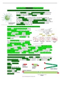
The centrosome is one example of such a cytoskeletal organiser: in
addition to nucleating the growth of microtubules, it holds them together
in a defined organisation, with the plus ends pointing outward. In this
way, the centrosome creates the astral array of microtubules that is able to
find the centre of each cell – this self-centring mechanism controls
transport polarity within the cell.
Actin filaments are organised into several types of array
Bundles and web-like (gel-like) networks. These
different structures are initiated by the action of
distinct nucleating proteins: the long straight
filaments produced by formins make tight parallel
bundles and the ARP complex makes dendritic
networks/ webs (binds to actin filaments and
promotes formation of a new filament at a 70
degree angle).
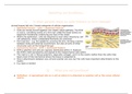
E.g. a fibroblast crawling (targeting a bacterium or
migrating during development) has structures
with different arrangements of actin filaments.
Filopodia are spike-like projections of the plasma
membrane that allow a cell to explore its environment – composed of tight bundles of actin
filaments organised parallel to each other. Lamellipodium is a dendritic branching structure
which pushes the cell forward. Behind this, the cell cortex has a gel-like network, involving
linkage and interaction between different filaments. Finally, stress fibres are formed of anti-
parallel bundles which are more widely spaced than in filopodia, and are responsible for
attaching to the substrate via integrins and contracting, pulling the rear of the cell forward.
Actin cross-linking proteins have two actin binding sites
Actin filament cross-linking proteins help to stabilise and maintain these distinct structures,
which is important due to the brittle nature of the filaments. Each of these proteins has two
actin-binding domains (red) which are related in sequence – each site links to one fibre, in
order to cross-link them.
4 types of actin binding
proteins:
Fimbrin has two directly
adjacent actin-binding sites,
so that it holds its two actin
filaments very close
together, aligned with the
same polarity.
, a-actinin has two actin-binding sites which are separated by a spacer (forms an anti-parallel
dimer), so that it forms more loosely packed actin bundles.
Filamin has two actin-binding sites with a V-shaped flexible linkage between them, so that it
cross-links actin filaments into a network with the filaments orientated almost at right angles
to one another.
Spectrin is a tetramer of two alpha and two beta subunits, and the tetramer has two actin-
binding sites. In the red blood cell, spectrin is concentrated just beneath the plasma
membrane, forming a network which creates a stiff cell cortex that provides mechanical
support for the PM, allowing the red blood cell to spring back to its original shape after
squeezing through a capillary.
Filamin and spectrin stabilise filament webs
Any cross-linking protein that has its two actin binding
domains joined by a long bent linkage can form a gel-like
mesh of filaments which provides mechanical strength.
Filamin promotes the formation of a loose and highly
viscous gel by clamping together two actin filaments
roughly at right angles. Cells require the actin gels formed
by filamin in order to extend the thin sheet-like
membrane projections called lamellipodia that help them
to crawl across solid surfaces. Filamin is lacking in some
types of cancer cells, especially some malignant
melanomas, which cannot crawl properly. Such cells are
less invasive than melanoma cells expressing filamin, and
so the cancer is less likely to metastasise.
Multiple filamin proteins have tissue-specific expression –
e.g. a mutation in filaminA gene causes significant
changes in brain structure due to migration problems.
Fimbrin and α-actinin form actin filament
bundles
Fimbrin is a small monomeric bundling protein which
excludes myosin, and so cross-links actin filaments into
tight parallel bundles which are not contractile. E.g.
microspikes, filopodia, and microvilli
a-actinin is a homodimer which cross-links actin
filaments into loose anti—parallel bundles , which allow
the motor protein myosin II to participate in the
assembly, making the filaments contractile. E.g. stress
fibres and muscle cells
Fimbrin and a-actinin tend to exclude one another
because of the very different spacing of the actin filament
bundles that they form. The two types of bundling
protein are themselves mutually exclusive.
PLectin (Plakin family) links the IF, MT and actin cytoskeleton
Cytoskeletal filaments are organised into higher-order structures in cells
For the cytoskeleton to form a useful intracellular scaffold that provides
the cell with mechanical integrity, determines its shape and ensures its
function, the filaments must be organised.
The centrosome is one example of such a cytoskeletal organiser: in
addition to nucleating the growth of microtubules, it holds them together
in a defined organisation, with the plus ends pointing outward. In this
way, the centrosome creates the astral array of microtubules that is able to
find the centre of each cell – this self-centring mechanism controls
transport polarity within the cell.
Actin filaments are organised into several types of array
Bundles and web-like (gel-like) networks. These
different structures are initiated by the action of
distinct nucleating proteins: the long straight
filaments produced by formins make tight parallel
bundles and the ARP complex makes dendritic
networks/ webs (binds to actin filaments and
promotes formation of a new filament at a 70
degree angle).
E.g. a fibroblast crawling (targeting a bacterium or
migrating during development) has structures
with different arrangements of actin filaments.
Filopodia are spike-like projections of the plasma
membrane that allow a cell to explore its environment – composed of tight bundles of actin
filaments organised parallel to each other. Lamellipodium is a dendritic branching structure
which pushes the cell forward. Behind this, the cell cortex has a gel-like network, involving
linkage and interaction between different filaments. Finally, stress fibres are formed of anti-
parallel bundles which are more widely spaced than in filopodia, and are responsible for
attaching to the substrate via integrins and contracting, pulling the rear of the cell forward.
Actin cross-linking proteins have two actin binding sites
Actin filament cross-linking proteins help to stabilise and maintain these distinct structures,
which is important due to the brittle nature of the filaments. Each of these proteins has two
actin-binding domains (red) which are related in sequence – each site links to one fibre, in
order to cross-link them.
4 types of actin binding
proteins:
Fimbrin has two directly
adjacent actin-binding sites,
so that it holds its two actin
filaments very close
together, aligned with the
same polarity.
, a-actinin has two actin-binding sites which are separated by a spacer (forms an anti-parallel
dimer), so that it forms more loosely packed actin bundles.
Filamin has two actin-binding sites with a V-shaped flexible linkage between them, so that it
cross-links actin filaments into a network with the filaments orientated almost at right angles
to one another.
Spectrin is a tetramer of two alpha and two beta subunits, and the tetramer has two actin-
binding sites. In the red blood cell, spectrin is concentrated just beneath the plasma
membrane, forming a network which creates a stiff cell cortex that provides mechanical
support for the PM, allowing the red blood cell to spring back to its original shape after
squeezing through a capillary.
Filamin and spectrin stabilise filament webs
Any cross-linking protein that has its two actin binding
domains joined by a long bent linkage can form a gel-like
mesh of filaments which provides mechanical strength.
Filamin promotes the formation of a loose and highly
viscous gel by clamping together two actin filaments
roughly at right angles. Cells require the actin gels formed
by filamin in order to extend the thin sheet-like
membrane projections called lamellipodia that help them
to crawl across solid surfaces. Filamin is lacking in some
types of cancer cells, especially some malignant
melanomas, which cannot crawl properly. Such cells are
less invasive than melanoma cells expressing filamin, and
so the cancer is less likely to metastasise.
Multiple filamin proteins have tissue-specific expression –
e.g. a mutation in filaminA gene causes significant
changes in brain structure due to migration problems.
Fimbrin and α-actinin form actin filament
bundles
Fimbrin is a small monomeric bundling protein which
excludes myosin, and so cross-links actin filaments into
tight parallel bundles which are not contractile. E.g.
microspikes, filopodia, and microvilli
a-actinin is a homodimer which cross-links actin
filaments into loose anti—parallel bundles , which allow
the motor protein myosin II to participate in the
assembly, making the filaments contractile. E.g. stress
fibres and muscle cells
Fimbrin and a-actinin tend to exclude one another
because of the very different spacing of the actin filament
bundles that they form. The two types of bundling
protein are themselves mutually exclusive.
PLectin (Plakin family) links the IF, MT and actin cytoskeleton