Chapter 5: Plasma membranes
5.1: structure and function of membranes
Membrane structure
All the membranes in a cell have the same basic structure.
The cell surface membrane which separates the cell from its external environment is known as the plasma
membrane.
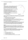
Membranes are formed from a phospholipid bilayer.
The hydrophilic phosphate heads of the phospholipids form both the inner and outer surface of a
membrane, sandwiching the fatty acid tails of the phospholipids to form a hydrophobic core inside the
membrane.
Cells normally exist in aqueous environments.
The inside of cells and organelles are also usually aqueous environments.
Phospholipid bilayers are perfectly suited as membranes because the outer surfaces of the hydrophilic
phosphate heads can interact with water.
Cell membrane theory
Membranes were seen for the first time following the invention of electron microscopy, which allowed
images to be taken with higher magnification and resolution.
Images taken in the 1950s showed the membrane as two black parallel lines - supporting an earlier theory
that membranes were composed of a lipid bilayer.
In 1972 American scientists Singer and Nicolson proposed a model. building upon an earlier lipid-bilayer
model, in which proteins occupy various positions in the membrane.
The model is known as the fluid- mosaic model because the phospholipids are free to move within
the layer relative to each other they are fluid, giving the membrane flexibility, and because the proteins
embedded in the bilayer vary in shape, size, and position in the same way as the tiles of a mosaic.
This model forms the basis of our understanding of membranes today.
Membrane proteins
Membrane proteins have important roles in the various functions of membranes. There are two types of
proteins in the cell-surface membrane - intrinsic and extrinsic proteins.
Intrinsic proteins
Intrinsic proteins, or integral proteins, are transmembrane proteins that are embedded through both layers
of a membrane.
They have amino acids with hydrophobic R-groups on their external surfaces, which interact with the
hydrophobic core of the membrane, keeping them in place.
Channel and carrier proteins are intrinsic proteins.
They are both involved in transport across the membrane.
Channel proteins provide a hydrophilic channel that allows the passive movement of polar molecules and
ions down a concentration gradient through membranes.
They are held in position by interactions between the hydrophobic core of the membrane and the
hydrophobic R-groups on the outside of the proteins.
Carrier proteins have an important role in both passive transport and active transport into cells
This often involves the shape of the protein changing.
Glycoproteins
Glycoproteins are intrinsic proteins.
They are embedded in the cell-surface membrane with attached carbohydrate chains of varying lengths and
shapes.
Glycoproteins play a role in cell adhesion when cells join together to form tight junctions in certain tissues,
and as receptors for chemical signals.
, When the chemical binds to the receptor, it elicits a response from the cell.
This may cause a direct response or set off a cascade of events inside the cell.
This process is known as cell communication or cell signalling.
Examples include:
o receptors for neurotransmitters such as acetylcholine at nerve cell synapses.
o The binding of the neurotransmitters triggers or prevents an impulse in the next neurone receptors
for peptide hormones, including insulin and glucagon, which affect the uptake and storage of glucose
by cells.
Some drugs act by binding to cell receptors.
o For example, 3 blockers are used to reduce the response of the heart to stress.
Glycolipids
Glycolipids are like glycoproteins. They are lipids with attached carbohydrate (sugar) chains.
These molecules are called cell markers or antigens and can be recognised by the cells of the immune system
as self (of the organism) or non-self
Extrinsic proteins
Extrinsic proteins or peripheral proteins are present in one side of the bilayer.
They normally have hydrophilic R-groups on their outer surfaces and interact with the polar heads of the
phospholipids or with intrinsic proteins.
They can be present in either layer or some move between layers.
Cholesterol
Cholesterol is a lipid with a hydrophilic end and a hydrophobic end, like a phospholipid.
It regulates the fluidity of membranes.
Cholesterol molecules are positioned between phospholipids in a membrane bilayer, with the hydrophilic
end interacting with the heads and the hydrophobic end interacting with the tails, pulling
them together.
In this way cholesterol adds stability to membranes without making them too rigid.
The cholesterol molecules prevent the membranes becoming too solid by stopping the phospholipid
molecules from grouping too closely and crystallising.
Sites of chemical reactions
Like enzymes, proteins in the membranes forming organelles, or present within organelles, have to be in
particular positions for chemical reactions to take place.
For example, the electron carriers and the enzyme ATP synthase must be in the correct positions
within the cristae (inner membrane of mitochondrion) to produce ATP in respiration.
The enzymes of photosynthesis are found on the membrane stacks within the chloroplasts.
5.2: Factors affecting membrane structures
Temperature
Phospholipids in a cell membrane are constantly moving.
When temperature is increased the phospholipids will have more kinetic energy and will move more.
This makes a membrane more fluid, and it begins to lose its structure.
If temperature continues to increase the cell will eventually break down completely.
This loss of structure increases the permeability of the membrane, making it easier for particles to cross it.
Carrier and channel proteins in the membrane will be denatured at higher temperatures.
These proteins are involved in transport across the membrane so as they denature, membrane permeability
will be affected.
Solvents
5.1: structure and function of membranes
Membrane structure
All the membranes in a cell have the same basic structure.
The cell surface membrane which separates the cell from its external environment is known as the plasma
membrane.
Membranes are formed from a phospholipid bilayer.
The hydrophilic phosphate heads of the phospholipids form both the inner and outer surface of a
membrane, sandwiching the fatty acid tails of the phospholipids to form a hydrophobic core inside the
membrane.
Cells normally exist in aqueous environments.
The inside of cells and organelles are also usually aqueous environments.
Phospholipid bilayers are perfectly suited as membranes because the outer surfaces of the hydrophilic
phosphate heads can interact with water.
Cell membrane theory
Membranes were seen for the first time following the invention of electron microscopy, which allowed
images to be taken with higher magnification and resolution.
Images taken in the 1950s showed the membrane as two black parallel lines - supporting an earlier theory
that membranes were composed of a lipid bilayer.
In 1972 American scientists Singer and Nicolson proposed a model. building upon an earlier lipid-bilayer
model, in which proteins occupy various positions in the membrane.
The model is known as the fluid- mosaic model because the phospholipids are free to move within
the layer relative to each other they are fluid, giving the membrane flexibility, and because the proteins
embedded in the bilayer vary in shape, size, and position in the same way as the tiles of a mosaic.
This model forms the basis of our understanding of membranes today.
Membrane proteins
Membrane proteins have important roles in the various functions of membranes. There are two types of
proteins in the cell-surface membrane - intrinsic and extrinsic proteins.
Intrinsic proteins
Intrinsic proteins, or integral proteins, are transmembrane proteins that are embedded through both layers
of a membrane.
They have amino acids with hydrophobic R-groups on their external surfaces, which interact with the
hydrophobic core of the membrane, keeping them in place.
Channel and carrier proteins are intrinsic proteins.
They are both involved in transport across the membrane.
Channel proteins provide a hydrophilic channel that allows the passive movement of polar molecules and
ions down a concentration gradient through membranes.
They are held in position by interactions between the hydrophobic core of the membrane and the
hydrophobic R-groups on the outside of the proteins.
Carrier proteins have an important role in both passive transport and active transport into cells
This often involves the shape of the protein changing.
Glycoproteins
Glycoproteins are intrinsic proteins.
They are embedded in the cell-surface membrane with attached carbohydrate chains of varying lengths and
shapes.
Glycoproteins play a role in cell adhesion when cells join together to form tight junctions in certain tissues,
and as receptors for chemical signals.
, When the chemical binds to the receptor, it elicits a response from the cell.
This may cause a direct response or set off a cascade of events inside the cell.
This process is known as cell communication or cell signalling.
Examples include:
o receptors for neurotransmitters such as acetylcholine at nerve cell synapses.
o The binding of the neurotransmitters triggers or prevents an impulse in the next neurone receptors
for peptide hormones, including insulin and glucagon, which affect the uptake and storage of glucose
by cells.
Some drugs act by binding to cell receptors.
o For example, 3 blockers are used to reduce the response of the heart to stress.
Glycolipids
Glycolipids are like glycoproteins. They are lipids with attached carbohydrate (sugar) chains.
These molecules are called cell markers or antigens and can be recognised by the cells of the immune system
as self (of the organism) or non-self
Extrinsic proteins
Extrinsic proteins or peripheral proteins are present in one side of the bilayer.
They normally have hydrophilic R-groups on their outer surfaces and interact with the polar heads of the
phospholipids or with intrinsic proteins.
They can be present in either layer or some move between layers.
Cholesterol
Cholesterol is a lipid with a hydrophilic end and a hydrophobic end, like a phospholipid.
It regulates the fluidity of membranes.
Cholesterol molecules are positioned between phospholipids in a membrane bilayer, with the hydrophilic
end interacting with the heads and the hydrophobic end interacting with the tails, pulling
them together.
In this way cholesterol adds stability to membranes without making them too rigid.
The cholesterol molecules prevent the membranes becoming too solid by stopping the phospholipid
molecules from grouping too closely and crystallising.
Sites of chemical reactions
Like enzymes, proteins in the membranes forming organelles, or present within organelles, have to be in
particular positions for chemical reactions to take place.
For example, the electron carriers and the enzyme ATP synthase must be in the correct positions
within the cristae (inner membrane of mitochondrion) to produce ATP in respiration.
The enzymes of photosynthesis are found on the membrane stacks within the chloroplasts.
5.2: Factors affecting membrane structures
Temperature
Phospholipids in a cell membrane are constantly moving.
When temperature is increased the phospholipids will have more kinetic energy and will move more.
This makes a membrane more fluid, and it begins to lose its structure.
If temperature continues to increase the cell will eventually break down completely.
This loss of structure increases the permeability of the membrane, making it easier for particles to cross it.
Carrier and channel proteins in the membrane will be denatured at higher temperatures.
These proteins are involved in transport across the membrane so as they denature, membrane permeability
will be affected.
Solvents