Kidney
P.393. Histology class, look for:
● Parietal epithelial cell
● Bowman’s capsule
● Proximal tubule
● Distal tubule
● Arteria arcuata (arcuate artery, on the border of cortex/medulla)
● Glomerulus (vascular pole/tubular pole)
○ Endothelial cell
○ Podocyte
○ Mesangial cell
Figures below were collected from the book Junqueira's "Basic Histology Text and Atlas", 14th edition, by
Anthony L. Mescher:
Figure 19-5 Renal corpuscles
(a)The renal corpuscle is a small mass of capillaries called the glomerulus housed within a bulbous glomerular
capsule. The internal lining of the capsule is composed of complex epithelial cells called podocytes, which cover
each capillary, forming slit-like spaces between interdigitating processes called pedicels. Blood enters and
leaves the glomerulus through the afferent and efferent arterioles, respectively.
(b)The micrograph shows the major histologic features of a renal corpuscle. The glomerulus (G) of capillaries is
surrounded by the capsular space (CS) covered by the simple squamous parietal layer (PL) of Bowman capsule.
Near the corpuscle is that nephron’s macula densa (MD) and sections of proximal convoluted tubules (PCT) and
distal convoluted tubules (DCT). (H&E; X300)
, Figure 19-7 Mesangium
a) Diagram shows that mesangial cells in renal corpuscles are located between capillaries and cover those
capillary surface not covered by podocyte processes.
Figure 19-8 Renal cortex: proximal and distal convoluted tubules
a) The micrograph shows the continuity at a renal corpuscle’s tubular pole (TP) between the simple cuboidal
epithelium of a proximal convoluted tubule (P) and the simple squamous epithelium of the capsule’s parietal
layer. The urinary space (U) between the parietal layer and the glomerulus (G) drains into the lumen of the
proximal tubule. The lumens of the proximal tubules appear filled, because of the long microvilli of the brush
border and aggregates of small plasma proteins bound to this structure. By contrast, the lumens of distal
convoluted tubules (D) appear empty, lacking a brush border and protein.
(b) Here the abundant peritubular capillaries and draining venules (arrows) that surround the proximal (P) and
distal (D) convoluted tubules are clearly seen. (Both X400; H&E)
, Figure 19-9 Convoluted tubules, nephron loops and collecting ducts
b) A section of cortical tissue shows one renal corpuscle (RC), the wide, eosinophilic proximal convoluted tubules
(PCT) with the smaller, less well-stained distal convoluted tubules (DCT). (X160; H&E)
(c) Diagram shows the major structural differences between the cuboidal cells of proximal and distal tubules.
Cells of both tubules have basal membrane invaginations associated with mitochondria.
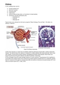
Represented below on Image 1, the Glomerulus can be recognised as it stands out from the rest of the
tissue due to its structure: the basement membrane on the Bowman’s capsule (surrounding the glomerulus) is
composed of simple squamous epithelium (parietal epithelium), there is an open space called Bowman's space
and in the middle there are capillary loops, surrounded by podocytes and mesangial cells, all three together work
as a renal filtration apparatus 1. The Glomerulus is also always attached to an arteriole (vascular pole) and has
an opening in the Bowman's capsule to a proximal tubule (urinary pole). Podocytes are star-shaped epithelial
cells and mesangial cells hold together the capillaries, both are are about the same size and hard to differentiate
from each other, but the mesangial cells are usually stained more darkly1. The capillaries are lined with
endothelial cells can be identified by lighter colour of their stain and the common presence of one or two
erythrocytes. The glomeruli's function is to filtrate the blood plasma in the capillaries into a "glomerular filtrate",
which will become urine 1.
Image 1. Graphic representation of a glomeruli structure: Bowman's capsule and space and capillary loops
(podocytes, mesangial and endothelial cells) – kidney tissue, HE stain (40X)
Next to the previously mentioned tubular pole of the glomerulus is the proximal convoluted tubule (Image
2), which can be identified by its simple cuboidal epithelium, acidophillic/eosinophilic (pink) cytoplasm, big
(purple) nuclei, a microvilli brush border and the appearance of always being filled with filtrate, that moves
towards the loop of Henle1. In comparison, the distal convoluted tubules (Image 3) can be identified by their
smaller size, flatter cells, with a bigger cytoplasm to nucleus ratio (said cytoplasm is less acidophillic/eosinophilic
so it appears in a lighter pink) and with no brush border, giving it the appearance of being empty. Both are
present in the cortex, PCT cells in reabsorption of all organic nutrients and proteins/most water and electrolytes,
P.393. Histology class, look for:
● Parietal epithelial cell
● Bowman’s capsule
● Proximal tubule
● Distal tubule
● Arteria arcuata (arcuate artery, on the border of cortex/medulla)
● Glomerulus (vascular pole/tubular pole)
○ Endothelial cell
○ Podocyte
○ Mesangial cell
Figures below were collected from the book Junqueira's "Basic Histology Text and Atlas", 14th edition, by
Anthony L. Mescher:
Figure 19-5 Renal corpuscles
(a)The renal corpuscle is a small mass of capillaries called the glomerulus housed within a bulbous glomerular
capsule. The internal lining of the capsule is composed of complex epithelial cells called podocytes, which cover
each capillary, forming slit-like spaces between interdigitating processes called pedicels. Blood enters and
leaves the glomerulus through the afferent and efferent arterioles, respectively.
(b)The micrograph shows the major histologic features of a renal corpuscle. The glomerulus (G) of capillaries is
surrounded by the capsular space (CS) covered by the simple squamous parietal layer (PL) of Bowman capsule.
Near the corpuscle is that nephron’s macula densa (MD) and sections of proximal convoluted tubules (PCT) and
distal convoluted tubules (DCT). (H&E; X300)
, Figure 19-7 Mesangium
a) Diagram shows that mesangial cells in renal corpuscles are located between capillaries and cover those
capillary surface not covered by podocyte processes.
Figure 19-8 Renal cortex: proximal and distal convoluted tubules
a) The micrograph shows the continuity at a renal corpuscle’s tubular pole (TP) between the simple cuboidal
epithelium of a proximal convoluted tubule (P) and the simple squamous epithelium of the capsule’s parietal
layer. The urinary space (U) between the parietal layer and the glomerulus (G) drains into the lumen of the
proximal tubule. The lumens of the proximal tubules appear filled, because of the long microvilli of the brush
border and aggregates of small plasma proteins bound to this structure. By contrast, the lumens of distal
convoluted tubules (D) appear empty, lacking a brush border and protein.
(b) Here the abundant peritubular capillaries and draining venules (arrows) that surround the proximal (P) and
distal (D) convoluted tubules are clearly seen. (Both X400; H&E)
, Figure 19-9 Convoluted tubules, nephron loops and collecting ducts
b) A section of cortical tissue shows one renal corpuscle (RC), the wide, eosinophilic proximal convoluted tubules
(PCT) with the smaller, less well-stained distal convoluted tubules (DCT). (X160; H&E)
(c) Diagram shows the major structural differences between the cuboidal cells of proximal and distal tubules.
Cells of both tubules have basal membrane invaginations associated with mitochondria.
Represented below on Image 1, the Glomerulus can be recognised as it stands out from the rest of the
tissue due to its structure: the basement membrane on the Bowman’s capsule (surrounding the glomerulus) is
composed of simple squamous epithelium (parietal epithelium), there is an open space called Bowman's space
and in the middle there are capillary loops, surrounded by podocytes and mesangial cells, all three together work
as a renal filtration apparatus 1. The Glomerulus is also always attached to an arteriole (vascular pole) and has
an opening in the Bowman's capsule to a proximal tubule (urinary pole). Podocytes are star-shaped epithelial
cells and mesangial cells hold together the capillaries, both are are about the same size and hard to differentiate
from each other, but the mesangial cells are usually stained more darkly1. The capillaries are lined with
endothelial cells can be identified by lighter colour of their stain and the common presence of one or two
erythrocytes. The glomeruli's function is to filtrate the blood plasma in the capillaries into a "glomerular filtrate",
which will become urine 1.
Image 1. Graphic representation of a glomeruli structure: Bowman's capsule and space and capillary loops
(podocytes, mesangial and endothelial cells) – kidney tissue, HE stain (40X)
Next to the previously mentioned tubular pole of the glomerulus is the proximal convoluted tubule (Image
2), which can be identified by its simple cuboidal epithelium, acidophillic/eosinophilic (pink) cytoplasm, big
(purple) nuclei, a microvilli brush border and the appearance of always being filled with filtrate, that moves
towards the loop of Henle1. In comparison, the distal convoluted tubules (Image 3) can be identified by their
smaller size, flatter cells, with a bigger cytoplasm to nucleus ratio (said cytoplasm is less acidophillic/eosinophilic
so it appears in a lighter pink) and with no brush border, giving it the appearance of being empty. Both are
present in the cortex, PCT cells in reabsorption of all organic nutrients and proteins/most water and electrolytes,