LEVELS OF BIOLOGICAL ORGANISATION
1. Organs and systems
2. Tissues
3. Cells
4. Organelles
5. Molecules
HIERARCHY OF CELLS
1. Subcellular (organelles)
2. Intracellular (everything within the membrane)
3. Molecules making up organelles
4. Molecules making up the environment (extracellular matrix)
5. Interaction of cellular molecules (intracellular or extracellular)
WHAT ARE CELLS?
● smallest unit of organisation that can perform all the activities required for life
● The organism’s basic units of structure and function.
What makes a cell?
- Enclosed in a membrane
- Use DNA as their genetic info
- Can divide - for reproduction, growth and repair
- Every cell arises from another cell
Activities performed by a cell
● Smallest unit that can carry out all life activities:
○ Extraction of energy from raw materials
1
, ○ Growth in an organized manner
○ Respond to stimuli
○ Homeostasis
○ Reproduce
○ Adapt
Importance of cell size
- Cells are small to make it easier to perform tasks. Smaller organisms can
complete the tasks more efficiently.
- Having many cells performing one task increases the efficiency and helps divide
up tasks
- The greater the number of cells in an area, the greater the surface area thus
increasing efficiency (many cells are working in that area at once)
- In the context of multicellular organisms, many cells result in a large surface
area of that organism. Thus many more reactions occurring at a time and often
in specialized compartments. (Think of a Rubick’s cube)
- The surface area to volume ratio is critical: as a cell increases in size its surface
area grows proportionately less than its volume.
Measuring cells
- Measured in μm (x10^-6)
- The angstrom is used to express atoms, molecules and other microscopic
structures (1 Angstrom = 1x10^-10)
DISCOVERY OF CELLS
- Robert Hooke discovered the first cell - using a microscope
- He coined the term cell because the structures were box-like
2
, - The structures he described were dead cork tissue of an oak bark.
- Antoni van Leeuwenhoek saw the first living cells
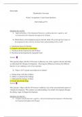
CELL THEORY (NB!)
1. All organisms are composed of one or more cells
2. Cells are the smallest living organisms
3. Cells arise only by division of a previously existing cell
MICROSCOPY
● Microscopy is the technical field of using microscopes to view objects and areas
of objects that cannot be seen with the naked eye.
The parameters of microscopy are:
1. Magnification: the ratio of an object’s image size to a real size
2. Resolution: a measure of the clarity of the image; the minimum distance two
points can be separated and still be distinguished as separate points
3. Contrast: The difference in brightness between the light and dark areas of an
image
Note: for the light microscope, light is also an important parameter.
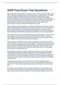
1. The light microscope
● Light microscope: visible light is used to illuminate the specimen. Light passes
through the lens and magnifies the image.
How it works:
- A compound light microscope has more than one lens
3
, - The light is shone from the bottom of the microscope and onto the specimen via
the condenser lens
- The light passes through the specimen and passes through the objective lens
(magnifying lens)
- The light then enters the projector lens
- The light is then directed to the ocular lens (the one you look through)
Therefore total magnification of the specimen is objective magnification X ocular lens
magnification.
μm
What can be seen:
- larger components of the cell such as nuclei, nucleoli, secretory granules,
lysosomes, and large mitochondria.
Staining techniques can be used to improve resolution/contrast. Disadvantageous
because it sometimes kills the cells.
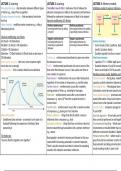
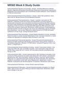
Types of light microscopy
1. Brightfield - Light passes through the specimen. Staining with dyes can enhance
the contrast. Disadvantageous as staining can kill the cells.
2. Fluorescence - UV light is shone through the specimen, and visible light is
emitted to create a visible image.
4
, 3. Phase-contrast - Enhances the contrast of unstained cells by amplifying
variations in density. Used for unpigmented, living cells.
4. Differential-interference-contrast (Nomarski) - uses optical modifications to
exaggerate differences in density, producing an almost 3D image
5. Confocal - another form of fluorescent microscopy - the specimen is sectioned,
several images are taken at different planes, and out of focus light is removed.
3D reconstruction produces a clear image
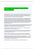
2. Electron Microscopy
- The specimen is flooded with fast-moving electrons instead of light
Types
● Scanning electron microscopy
- Focuses the electrons on the surface
- Gives you an image of the outside surface of the specimen
● Transmission electron microscopy
- Electrons are passed through the specimen
- Internal structures of the specimen are seen
How it works
- Similar to light microscope
- Light source is replaced by a beam of very fast moving electrons
- An electron micrograph is presented on a screen.
Disadvantages of electron microscopy
- The preparation of specimen renders the specimens date and kills it
Importance of microscopy
- Helps us understand cell structures
5
, - Can expose the different compartments of cells
- Helps biochemists link the structure of cells to their function
- It is important to use biochemical methods to help understand cellular structure
and function
- Cell structures can be correlated to cell function
CELLULAR FRACTIONATION
● The process used to separate cellular components while preserving individual
functions of each component
- helps scientist determine functions of organelles
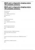
Centrifugation
● Technique that helps to separate mixtures by applying centrifugal force
● Cells can be separated according to their size and density using this method
● Cells/solution of cells are put into a test tube, broken up (via homegenation or
chemical methods), and spun around
● Heavier components collect at the bottom (called the pallet) and lighter pieces
collect at the top (called the supernatant)
PROKARYOTES AND EUKARYOTES
General features of all cells:
1. Contain chromosomes which carry genes in the form of DNA
2. Have ribosomes which make proteins
3. Bounded by a selective barrier - the plasma membrane/cell membrane
6
1. Organs and systems
2. Tissues
3. Cells
4. Organelles
5. Molecules
HIERARCHY OF CELLS
1. Subcellular (organelles)
2. Intracellular (everything within the membrane)
3. Molecules making up organelles
4. Molecules making up the environment (extracellular matrix)
5. Interaction of cellular molecules (intracellular or extracellular)
WHAT ARE CELLS?
● smallest unit of organisation that can perform all the activities required for life
● The organism’s basic units of structure and function.
What makes a cell?
- Enclosed in a membrane
- Use DNA as their genetic info
- Can divide - for reproduction, growth and repair
- Every cell arises from another cell
Activities performed by a cell
● Smallest unit that can carry out all life activities:
○ Extraction of energy from raw materials
1
, ○ Growth in an organized manner
○ Respond to stimuli
○ Homeostasis
○ Reproduce
○ Adapt
Importance of cell size
- Cells are small to make it easier to perform tasks. Smaller organisms can
complete the tasks more efficiently.
- Having many cells performing one task increases the efficiency and helps divide
up tasks
- The greater the number of cells in an area, the greater the surface area thus
increasing efficiency (many cells are working in that area at once)
- In the context of multicellular organisms, many cells result in a large surface
area of that organism. Thus many more reactions occurring at a time and often
in specialized compartments. (Think of a Rubick’s cube)
- The surface area to volume ratio is critical: as a cell increases in size its surface
area grows proportionately less than its volume.
Measuring cells
- Measured in μm (x10^-6)
- The angstrom is used to express atoms, molecules and other microscopic
structures (1 Angstrom = 1x10^-10)
DISCOVERY OF CELLS
- Robert Hooke discovered the first cell - using a microscope
- He coined the term cell because the structures were box-like
2
, - The structures he described were dead cork tissue of an oak bark.
- Antoni van Leeuwenhoek saw the first living cells
CELL THEORY (NB!)
1. All organisms are composed of one or more cells
2. Cells are the smallest living organisms
3. Cells arise only by division of a previously existing cell
MICROSCOPY
● Microscopy is the technical field of using microscopes to view objects and areas
of objects that cannot be seen with the naked eye.
The parameters of microscopy are:
1. Magnification: the ratio of an object’s image size to a real size
2. Resolution: a measure of the clarity of the image; the minimum distance two
points can be separated and still be distinguished as separate points
3. Contrast: The difference in brightness between the light and dark areas of an
image
Note: for the light microscope, light is also an important parameter.
1. The light microscope
● Light microscope: visible light is used to illuminate the specimen. Light passes
through the lens and magnifies the image.
How it works:
- A compound light microscope has more than one lens
3
, - The light is shone from the bottom of the microscope and onto the specimen via
the condenser lens
- The light passes through the specimen and passes through the objective lens
(magnifying lens)
- The light then enters the projector lens
- The light is then directed to the ocular lens (the one you look through)
Therefore total magnification of the specimen is objective magnification X ocular lens
magnification.
μm
What can be seen:
- larger components of the cell such as nuclei, nucleoli, secretory granules,
lysosomes, and large mitochondria.
Staining techniques can be used to improve resolution/contrast. Disadvantageous
because it sometimes kills the cells.
Types of light microscopy
1. Brightfield - Light passes through the specimen. Staining with dyes can enhance
the contrast. Disadvantageous as staining can kill the cells.
2. Fluorescence - UV light is shone through the specimen, and visible light is
emitted to create a visible image.
4
, 3. Phase-contrast - Enhances the contrast of unstained cells by amplifying
variations in density. Used for unpigmented, living cells.
4. Differential-interference-contrast (Nomarski) - uses optical modifications to
exaggerate differences in density, producing an almost 3D image
5. Confocal - another form of fluorescent microscopy - the specimen is sectioned,
several images are taken at different planes, and out of focus light is removed.
3D reconstruction produces a clear image
2. Electron Microscopy
- The specimen is flooded with fast-moving electrons instead of light
Types
● Scanning electron microscopy
- Focuses the electrons on the surface
- Gives you an image of the outside surface of the specimen
● Transmission electron microscopy
- Electrons are passed through the specimen
- Internal structures of the specimen are seen
How it works
- Similar to light microscope
- Light source is replaced by a beam of very fast moving electrons
- An electron micrograph is presented on a screen.
Disadvantages of electron microscopy
- The preparation of specimen renders the specimens date and kills it
Importance of microscopy
- Helps us understand cell structures
5
, - Can expose the different compartments of cells
- Helps biochemists link the structure of cells to their function
- It is important to use biochemical methods to help understand cellular structure
and function
- Cell structures can be correlated to cell function
CELLULAR FRACTIONATION
● The process used to separate cellular components while preserving individual
functions of each component
- helps scientist determine functions of organelles
Centrifugation
● Technique that helps to separate mixtures by applying centrifugal force
● Cells can be separated according to their size and density using this method
● Cells/solution of cells are put into a test tube, broken up (via homegenation or
chemical methods), and spun around
● Heavier components collect at the bottom (called the pallet) and lighter pieces
collect at the top (called the supernatant)
PROKARYOTES AND EUKARYOTES
General features of all cells:
1. Contain chromosomes which carry genes in the form of DNA
2. Have ribosomes which make proteins
3. Bounded by a selective barrier - the plasma membrane/cell membrane
6