Module 1: Early vision - Learning objectives
Describe the basic properties of the eye
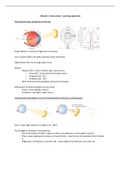
Pupil: dilates or restricts to light by iris (muscle)
Lens: projects light onto light-sensitive retina (inverted)
Signals leave the eye through optic nerve.
Retina:
- Macula (18°): central ‘yellow spot’: good vision.
o Fovea (5°): central area of sharpest vision.
o Parafovea (8 - 20°)
o Perifovea (18 - 20°)
- After that comes the periphery (not part of macula).
Distribution of photoreceptors across retina:
- Fovea: cones (details, colour)
- Periphery: rods (light, night vision)
Understand visual angle as a unit of measurement and why it is being used
Eye is a ball, light comes in at angle α (0 - 360°).
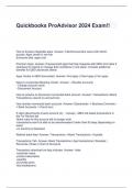
Visual angle as measure of eccentricity:
- Due to principles of optics: angle on retina corresponds to same angle in world.
- Thus: visual angle gives a measure of eccentricity – how far out into periphery the stimulus
is.
- Regardless of distance to observer (d) – same angle of eccentricity, but closer by.
,Visual angle as measure to express size of object’s projection:
- Here α expresses area on retina covered by the projection.
- This does change with distance! Distance d will change size of retinal projection & thus visual
angle.
- So objects far away seem smaller.
How far out can we see?
Explain the concept of a visual field
Visual field = part of the world that visual system responds to.
- In front of us.
- L/R visual fields cross in visual cortex.
Explain the concept of a receptive field
Receptive field = part of the world that a receptor/neuron responds to.
- Certain location in visual space
- Very small (high resolution) or very large (low resolution)
- Diff. receptors/neurons can have overlapping receptive fields.
- Convergence affects receptive field: spatial convergence is stronger for periphery →
receptive fields larger (also at cortical level stronger spatial convergence).
Draw out the pathway from eye to brain
, Note: nasal sides cross at the optic chiasm.
Explain what cortical magnification is
Cortical magnification = disproportionally large chunk of visual cortex (50%) is dedicated to the fovea
(< 1% of visual field).
- Peripheral vision gets less brain power (largely direct consequence of convergence & pooling
= receptive field size).
Explain the mechanism of lateral inhibition in center surround cells
Ganglion cell with ON centre-OFF surround receptive field:
- Mechanism behind supressing of the surround is called lateral inhibition.
- It likes local light in a dark surround better than local light in a light surround.
- Mechanism acts like Laplacian filter / DoG filter / Mexican hat filter: it increases local
contrast and sharpens the image (just like Photoshop).
Explain the more general idea of neurons acting as spatial filters
Visual neurons have special receptive fields, allowing them to act as spatial filters. You have centre-
surround cells, that help to sharpen an image. Then, extending the same lateral inhibition circuitry
Describe the basic properties of the eye
Pupil: dilates or restricts to light by iris (muscle)
Lens: projects light onto light-sensitive retina (inverted)
Signals leave the eye through optic nerve.
Retina:
- Macula (18°): central ‘yellow spot’: good vision.
o Fovea (5°): central area of sharpest vision.
o Parafovea (8 - 20°)
o Perifovea (18 - 20°)
- After that comes the periphery (not part of macula).
Distribution of photoreceptors across retina:
- Fovea: cones (details, colour)
- Periphery: rods (light, night vision)
Understand visual angle as a unit of measurement and why it is being used
Eye is a ball, light comes in at angle α (0 - 360°).
Visual angle as measure of eccentricity:
- Due to principles of optics: angle on retina corresponds to same angle in world.
- Thus: visual angle gives a measure of eccentricity – how far out into periphery the stimulus
is.
- Regardless of distance to observer (d) – same angle of eccentricity, but closer by.
,Visual angle as measure to express size of object’s projection:
- Here α expresses area on retina covered by the projection.
- This does change with distance! Distance d will change size of retinal projection & thus visual
angle.
- So objects far away seem smaller.
How far out can we see?
Explain the concept of a visual field
Visual field = part of the world that visual system responds to.
- In front of us.
- L/R visual fields cross in visual cortex.
Explain the concept of a receptive field
Receptive field = part of the world that a receptor/neuron responds to.
- Certain location in visual space
- Very small (high resolution) or very large (low resolution)
- Diff. receptors/neurons can have overlapping receptive fields.
- Convergence affects receptive field: spatial convergence is stronger for periphery →
receptive fields larger (also at cortical level stronger spatial convergence).
Draw out the pathway from eye to brain
, Note: nasal sides cross at the optic chiasm.
Explain what cortical magnification is
Cortical magnification = disproportionally large chunk of visual cortex (50%) is dedicated to the fovea
(< 1% of visual field).
- Peripheral vision gets less brain power (largely direct consequence of convergence & pooling
= receptive field size).
Explain the mechanism of lateral inhibition in center surround cells
Ganglion cell with ON centre-OFF surround receptive field:
- Mechanism behind supressing of the surround is called lateral inhibition.
- It likes local light in a dark surround better than local light in a light surround.
- Mechanism acts like Laplacian filter / DoG filter / Mexican hat filter: it increases local
contrast and sharpens the image (just like Photoshop).
Explain the more general idea of neurons acting as spatial filters
Visual neurons have special receptive fields, allowing them to act as spatial filters. You have centre-
surround cells, that help to sharpen an image. Then, extending the same lateral inhibition circuitry