Lecture Tractus Respiratorius
Anatomy of the lower airways
The lower airways consist of the conducting airways and the alveoli. The alveoli consist of
the alveolar sacs, ducts and respiratory bronchioles. Everything above the bronchioles are
the conducting airways.
Bronchoconstriction is the event in
which the trachea, bronchi and
intrapulmonary bronchi are constricting.
The trachea and main bronchi have a
cartilage layer around (horse shoe) and
smooth muscle contraction causes partial
closure of the airway, the airway cannot
close completely. Intrapulmonary
bronchi are the smallest bronchi and have
individual pieces of cartilage around for
support.
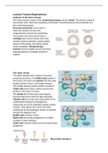
The upper airway
The upper airways have 2 types of mucosal
producing structures, the Goblet cells (yellow in
the picture, thin layer) and glands (the blue cells,
produce thicker mucus in the larger airways).
The smaller airways (bottom picture) consist of
Clubb cells (blue cells in bottom picture) that
produce a thin layer of mucus.
The alveoli do not have any mucus layers,
because that would inhibit the gas exchange.
Ciliated cells are small cells responsible for the
unidirectional transport of endogenous
compounds out of the respiratory system (pollen,
viruses). They deliver the mucus and other
compounds to the esophagus where it is cleared
through the digestive tract.
Goblet cells produce mucus.
Basal cells are airway progenitors (in smaller
numbers in lower airways).
Clubb cells secrete proteins and are small airway
progenitors.
Mucociliary transport
, When chloride is transported across the lumen, outside of the cell, an osmotic gradient is
formed, which attracts water. To compensate for the ion change, there are ion transporters
at the basal side. When there are defects in the ion channels, CFTR (cystic fibrosis) bay
occurs due to the imbalance in water.
Alveolar epithelial cells
Alveoli have surfactant within to prevent them from collapsing during exhalation. Type 1
cells in the alveoli regulate the gas-exchange. Type 2 cells are the surfactant progenitors.
Diseases of the respiratory tract:
- asthma
- Allergic rhinitis
- Cough
- COPD
- Pulmonary fibrosis
- Cystic fibrosis
Asthma is a heterogeneous disease, caused by an inflammation in the airways. Asthmatic
patients usually also have allergies due to airway hyperresponsiveness. The causes of
asthma include smoking and the hygiene hypothesis (we are born in a clean environment,
so as a baby we do not get in contact with diseases. This causes a more sensitive airway to
non dangerous compounds). There is no strong connection between hereditary genes and
asthma.
The inflammation is caused by the activation of mast cells, which release histamine that
causes the airway narrowing. This is triggered by cold and exercise e.g. An allergen binds to
the CD4 T-cell which
activates the Th0 cell to
becoming Th1 and Th2.
The Th2 cells release
interleukin factors that
activate eosinophils and
the production of B and P
cells. The IgE antibodies
from the B and P cells
then activate mast cells.
When there is a
continuous stimulus on the airway which leads to injury of the airway, the epithelium cannot
repair itself fast enough -> fibrosis.
Early allergic response IgE and mast-cell dependent. The late response is due to the
eosinophils that secrete various factors into the lung that lead to tissue damage.
Bronchospasms are a characteristic of the early phase and inflammation of the later phase.
Allergens lead to IgE production, then receptor signalling that causes the release of granules
with histamine. Histamine causes bronchoconstriction. Arachidonic acid is also formed upon
allergen contact, which leads to the production of prostaglandins and leukotrienes. These
factors also cause bronchoconstriction.
The parasympathetic nervous system (PNS) is also involved in the bronchoconstriction (that
is why allergic symptoms are worse during the night). The airway cholinergic system gets
Anatomy of the lower airways
The lower airways consist of the conducting airways and the alveoli. The alveoli consist of
the alveolar sacs, ducts and respiratory bronchioles. Everything above the bronchioles are
the conducting airways.
Bronchoconstriction is the event in
which the trachea, bronchi and
intrapulmonary bronchi are constricting.
The trachea and main bronchi have a
cartilage layer around (horse shoe) and
smooth muscle contraction causes partial
closure of the airway, the airway cannot
close completely. Intrapulmonary
bronchi are the smallest bronchi and have
individual pieces of cartilage around for
support.
The upper airway
The upper airways have 2 types of mucosal
producing structures, the Goblet cells (yellow in
the picture, thin layer) and glands (the blue cells,
produce thicker mucus in the larger airways).
The smaller airways (bottom picture) consist of
Clubb cells (blue cells in bottom picture) that
produce a thin layer of mucus.
The alveoli do not have any mucus layers,
because that would inhibit the gas exchange.
Ciliated cells are small cells responsible for the
unidirectional transport of endogenous
compounds out of the respiratory system (pollen,
viruses). They deliver the mucus and other
compounds to the esophagus where it is cleared
through the digestive tract.
Goblet cells produce mucus.
Basal cells are airway progenitors (in smaller
numbers in lower airways).
Clubb cells secrete proteins and are small airway
progenitors.
Mucociliary transport
, When chloride is transported across the lumen, outside of the cell, an osmotic gradient is
formed, which attracts water. To compensate for the ion change, there are ion transporters
at the basal side. When there are defects in the ion channels, CFTR (cystic fibrosis) bay
occurs due to the imbalance in water.
Alveolar epithelial cells
Alveoli have surfactant within to prevent them from collapsing during exhalation. Type 1
cells in the alveoli regulate the gas-exchange. Type 2 cells are the surfactant progenitors.
Diseases of the respiratory tract:
- asthma
- Allergic rhinitis
- Cough
- COPD
- Pulmonary fibrosis
- Cystic fibrosis
Asthma is a heterogeneous disease, caused by an inflammation in the airways. Asthmatic
patients usually also have allergies due to airway hyperresponsiveness. The causes of
asthma include smoking and the hygiene hypothesis (we are born in a clean environment,
so as a baby we do not get in contact with diseases. This causes a more sensitive airway to
non dangerous compounds). There is no strong connection between hereditary genes and
asthma.
The inflammation is caused by the activation of mast cells, which release histamine that
causes the airway narrowing. This is triggered by cold and exercise e.g. An allergen binds to
the CD4 T-cell which
activates the Th0 cell to
becoming Th1 and Th2.
The Th2 cells release
interleukin factors that
activate eosinophils and
the production of B and P
cells. The IgE antibodies
from the B and P cells
then activate mast cells.
When there is a
continuous stimulus on the airway which leads to injury of the airway, the epithelium cannot
repair itself fast enough -> fibrosis.
Early allergic response IgE and mast-cell dependent. The late response is due to the
eosinophils that secrete various factors into the lung that lead to tissue damage.
Bronchospasms are a characteristic of the early phase and inflammation of the later phase.
Allergens lead to IgE production, then receptor signalling that causes the release of granules
with histamine. Histamine causes bronchoconstriction. Arachidonic acid is also formed upon
allergen contact, which leads to the production of prostaglandins and leukotrienes. These
factors also cause bronchoconstriction.
The parasympathetic nervous system (PNS) is also involved in the bronchoconstriction (that
is why allergic symptoms are worse during the night). The airway cholinergic system gets