Lecture 5: Bone Development
Learning Objectives
1) Identify the two types of bone growth
2) Explain the processes of bone development and bone healing
Types of Bone Growth
1. Intramembranous ossification
2. Endochondral ossification
NB: Ossification = bone formation
1. Intramembranous Ossification (mesenchymal template)
● New bone is formed from a template made of mesenchymal cells
○ This process involves the transformation of connective tissue
● Flat bones are formed by intramembranous ossification
● NB: Mesenchymal cells are multipotent stem cells that can be found in
the bone marrow that are capable of forming skeletal tissue
2. Endochondral (chondral = cartilage) Ossification (cartilaginous
template)
● New bone is formed from a cartilage template
● Most bones in the body form in this way
● Responsible for growth in bone length and forms the articular surfaces
○ Generates all three major areas of long bones: diaphysis,
epiphysis and metaphysis
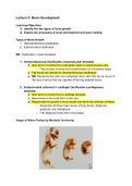
○ The diaphysis forms first in the foetus
Image of Kitten Foetus by Murdoch University
, Embryo stained with Alizarin Red
● Type of stain which shows calcium
● Alizarin Red will bind to the calcium in the developed bone
Primary Ossification in Kitten Embryo
● First areas (central bone - diaphysis) where bone is ossified - diaphysis first,
not epiphysis
● End of bones are not stained red as they have not yet ossified - these bones
ossify later in secondary ossification
By birth, most of the cartilage skeleton has been replaced with bone:
● Some areas of the cartilage model remain post-partum to allow for continued
bone growth through to adulthood when skeletal maturity is achieved
● The cartilage regions that remain in developing long bones in animals are
known as the growth plate (epiphyseal plate)
Learning Objectives
1) Identify the two types of bone growth
2) Explain the processes of bone development and bone healing
Types of Bone Growth
1. Intramembranous ossification
2. Endochondral ossification
NB: Ossification = bone formation
1. Intramembranous Ossification (mesenchymal template)
● New bone is formed from a template made of mesenchymal cells
○ This process involves the transformation of connective tissue
● Flat bones are formed by intramembranous ossification
● NB: Mesenchymal cells are multipotent stem cells that can be found in
the bone marrow that are capable of forming skeletal tissue
2. Endochondral (chondral = cartilage) Ossification (cartilaginous
template)
● New bone is formed from a cartilage template
● Most bones in the body form in this way
● Responsible for growth in bone length and forms the articular surfaces
○ Generates all three major areas of long bones: diaphysis,
epiphysis and metaphysis
○ The diaphysis forms first in the foetus
Image of Kitten Foetus by Murdoch University
, Embryo stained with Alizarin Red
● Type of stain which shows calcium
● Alizarin Red will bind to the calcium in the developed bone
Primary Ossification in Kitten Embryo
● First areas (central bone - diaphysis) where bone is ossified - diaphysis first,
not epiphysis
● End of bones are not stained red as they have not yet ossified - these bones
ossify later in secondary ossification
By birth, most of the cartilage skeleton has been replaced with bone:
● Some areas of the cartilage model remain post-partum to allow for continued
bone growth through to adulthood when skeletal maturity is achieved
● The cartilage regions that remain in developing long bones in animals are
known as the growth plate (epiphyseal plate)