Introduction
First, mouse muscle tissues are stained with haematoxylin and
eosin to obtain knowledge on essential steps in staining
procedures and on histology of muscle tissue. Haematoxylin is a
basic dye that stains DNA, while eosin is an acidic dye that stains
cytoplasmic proteins. After this, a different set of muscle tissues
is incubated with fast myosin heavy chain antibody. This
polyclonal primary antibody is produced in a rabbit. All
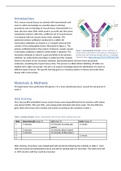
mammalian antibodies are based on a Y-shaped molecule that
consists of four polypeptide chains, illustrated by figure 1. The
primary antibody binds to the protein of interest, namely myosin.
Figure 1: Imunoglobulin structure. Source: Hofmann, A.
A secondary antibody is added to which biotin is attached. The (2018). Wilson and Walker’s Principles and Techniques of
secondary antibody is raised in a goat and binds to the primary Biochemistry and Molecular Biology (8th ed.). Cambridge
antibody. An avidin-biotin-peroxidase is added and this complex University Press. https://doi.org/10.1017/9781316677056
binds to the biotin on the secondary antibody. Diaminobenzidine will stain these peroxidase
molecules, visualizing the myosin heavy chain. This process is called indirect labelling. All slides are
studied with a light microscope. The aim is to acquire knowledge about the distribution of myosin in
different types of tissue. The specific learning goal is to visualize proteins in tissues and study these
tissues with a microscope.
Materials & Methods
All experiments were performed with gloves. For a more detailed protocol, consult the lab journal of
week 3.
H&E staining
First, five paraffin embedded mouse muscle tissues were deparaffinized by the teachers with xylene
and alcohol (100%, 96% and 70%), and subsequently hydrated with demi water. The five different
glass slides with tissue were marked and stained according to the schedule in table 1.
Table 1: Staining schedule. Mayer’s haematoxilin and 1% eosin (H&E) staining were used.
Slide Haematoxylin (step 1) H20 (step 2) Eosin (step 3)
1 5 minutes 10 minutes (running tap 1 minute
water)
2 5 minutes 10 minutes (demi water)
3 5 minutes 10 minutes (miliQ water)
4 1 minute
5
After staining, all sections were dehydrated with alcohol by following the schedule in table 2. Each
slide was stained and dehydrated one by one before starting with the next slide. The slides were left
in 100% alcohol until they could be mounted.
, Table 2: Dehydration schedule.
Step Alcohol (percentage) Time (seconds)
1 70% 10
2 80% 10
3 90% 10
4 96% 1
5 100% 2
6 100% 2
After dehydration, all sections were cleared with xylene and then a droplet of DePeX was added.
DePeX is a xylene based mounting medium. Each tissue section was covered with a cover slip. Lastly,
the sections were dried in a fume hood to remove xylene and subsequently sealed with nail polish.
The mouse muscle tissues could now be studied with a light microscope (Olympus).
Diaminobenzidine staining
There were four slides with 3 types of mouse muscle tissue on each slide; gastrocnemius, soleus, and
extensor digitorum longus. These four slides also needed to be deparaffinized first; this was done by
the teachers with xylene and 100% alcohol. Slides were kept in 100% alcohol to prevent them from
drying out. Table 3 demonstrates the first part of the immunohistochemical schedule of the glass
slides with muscle tissues. All these steps were the same for each slide and performed at room
temperature. Tris buffered saline (TBS) was used for all washing steps in the entire experiment.
Blocking with goat serum (step 5) needed to occur in a humidifying box.
Table 3: Immunohistochemical schedule. Methanol and hydrogen peroxide removes endogenous peroxidase, glycine blocks
free aldehyde groups, blocking with goat serum to prevent nonspecific binding of secondary antibody.
Step Solution Time (minutes) Notes
1 Methanol + 3% H2O2 30 20ml 30% H2O2 + 18ml ethanol
2 TBS (0.01M) 5x5 Washing step
3 TBS-glycine (0.01M) 30 1.51g glycine + 200ml 1x TBS
4 TBS (0.01M) 5x5 Washing step
5 5% goat serum 30 100 µl
(blocking buffer)
The slides were left in the incubation box during this part. Further incubations differed per slide. The
goat serum was tapped of from slides 1 and 2. Slide 1 was incubated with 100 µl fast myosin heavy
chain antibody (1/10000, rabbit) as primary antibody, slide 2 was incubated with 100 µl IgG (1/10000
in TBS/BSAc, rabbit). For slides 3 and 4, the goat serum from the blocking step could be left on the
tissue. Subsequently, all tissue sections were incubated overnight at 4 oC.
After the overnight incubation, all slides were washed with TBS (0.01M). A secondary antibody was
diluted to 1/200 by adding 5 µl biotinylated antibody (goat, anti-rabbit) to 995 µl TBS- BSAc
(Acetylated bovine serum). 100 µl of secondary antibody solution was then added to slides 1 and 2 in
First, mouse muscle tissues are stained with haematoxylin and
eosin to obtain knowledge on essential steps in staining
procedures and on histology of muscle tissue. Haematoxylin is a
basic dye that stains DNA, while eosin is an acidic dye that stains
cytoplasmic proteins. After this, a different set of muscle tissues
is incubated with fast myosin heavy chain antibody. This
polyclonal primary antibody is produced in a rabbit. All
mammalian antibodies are based on a Y-shaped molecule that
consists of four polypeptide chains, illustrated by figure 1. The
primary antibody binds to the protein of interest, namely myosin.
Figure 1: Imunoglobulin structure. Source: Hofmann, A.
A secondary antibody is added to which biotin is attached. The (2018). Wilson and Walker’s Principles and Techniques of
secondary antibody is raised in a goat and binds to the primary Biochemistry and Molecular Biology (8th ed.). Cambridge
antibody. An avidin-biotin-peroxidase is added and this complex University Press. https://doi.org/10.1017/9781316677056
binds to the biotin on the secondary antibody. Diaminobenzidine will stain these peroxidase
molecules, visualizing the myosin heavy chain. This process is called indirect labelling. All slides are
studied with a light microscope. The aim is to acquire knowledge about the distribution of myosin in
different types of tissue. The specific learning goal is to visualize proteins in tissues and study these
tissues with a microscope.
Materials & Methods
All experiments were performed with gloves. For a more detailed protocol, consult the lab journal of
week 3.
H&E staining
First, five paraffin embedded mouse muscle tissues were deparaffinized by the teachers with xylene
and alcohol (100%, 96% and 70%), and subsequently hydrated with demi water. The five different
glass slides with tissue were marked and stained according to the schedule in table 1.
Table 1: Staining schedule. Mayer’s haematoxilin and 1% eosin (H&E) staining were used.
Slide Haematoxylin (step 1) H20 (step 2) Eosin (step 3)
1 5 minutes 10 minutes (running tap 1 minute
water)
2 5 minutes 10 minutes (demi water)
3 5 minutes 10 minutes (miliQ water)
4 1 minute
5
After staining, all sections were dehydrated with alcohol by following the schedule in table 2. Each
slide was stained and dehydrated one by one before starting with the next slide. The slides were left
in 100% alcohol until they could be mounted.
, Table 2: Dehydration schedule.
Step Alcohol (percentage) Time (seconds)
1 70% 10
2 80% 10
3 90% 10
4 96% 1
5 100% 2
6 100% 2
After dehydration, all sections were cleared with xylene and then a droplet of DePeX was added.
DePeX is a xylene based mounting medium. Each tissue section was covered with a cover slip. Lastly,
the sections were dried in a fume hood to remove xylene and subsequently sealed with nail polish.
The mouse muscle tissues could now be studied with a light microscope (Olympus).
Diaminobenzidine staining
There were four slides with 3 types of mouse muscle tissue on each slide; gastrocnemius, soleus, and
extensor digitorum longus. These four slides also needed to be deparaffinized first; this was done by
the teachers with xylene and 100% alcohol. Slides were kept in 100% alcohol to prevent them from
drying out. Table 3 demonstrates the first part of the immunohistochemical schedule of the glass
slides with muscle tissues. All these steps were the same for each slide and performed at room
temperature. Tris buffered saline (TBS) was used for all washing steps in the entire experiment.
Blocking with goat serum (step 5) needed to occur in a humidifying box.
Table 3: Immunohistochemical schedule. Methanol and hydrogen peroxide removes endogenous peroxidase, glycine blocks
free aldehyde groups, blocking with goat serum to prevent nonspecific binding of secondary antibody.
Step Solution Time (minutes) Notes
1 Methanol + 3% H2O2 30 20ml 30% H2O2 + 18ml ethanol
2 TBS (0.01M) 5x5 Washing step
3 TBS-glycine (0.01M) 30 1.51g glycine + 200ml 1x TBS
4 TBS (0.01M) 5x5 Washing step
5 5% goat serum 30 100 µl
(blocking buffer)
The slides were left in the incubation box during this part. Further incubations differed per slide. The
goat serum was tapped of from slides 1 and 2. Slide 1 was incubated with 100 µl fast myosin heavy
chain antibody (1/10000, rabbit) as primary antibody, slide 2 was incubated with 100 µl IgG (1/10000
in TBS/BSAc, rabbit). For slides 3 and 4, the goat serum from the blocking step could be left on the
tissue. Subsequently, all tissue sections were incubated overnight at 4 oC.
After the overnight incubation, all slides were washed with TBS (0.01M). A secondary antibody was
diluted to 1/200 by adding 5 µl biotinylated antibody (goat, anti-rabbit) to 995 µl TBS- BSAc
(Acetylated bovine serum). 100 µl of secondary antibody solution was then added to slides 1 and 2 in