HC 01: Light and color
Overview & why different microscopes:
Choose microscope to match length scale, sample state (live/fixed), contrast needs, and temporal resolution.
Light microscopy:
• Strengths: live-cell imaging, molecular specificity (fluorophores/GFP), simpler sample prep, color.
• Limitations: diffraction-limited (~200 nm lateral), depth-of-field and out-of-focus blur in thick samples.
Electron microscopy (TEM/SEM):
• Strengths: nanometer/sub-nanometer resolution (ultrastructure).
• Limitations: requires fixation/dehydration/metal staining/coating; vacuum; no live imaging.
Decision checklist: live vs fixed; need molecular specificity; spatial scale; sample thickness; temporal
resolution.
Example: mitochondrial dynamics in live neurons → fluorescent tagging (GFP) + confocal or spinning-disk for
optical sectioning and reduced photodamage.
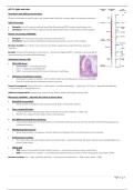
Histological staining: H&E
• What H&E shows:
o Hematoxylin → nuclei (blue/purple).
o Eosin → cytoplasm and ECM (pink).
o H&E = routine gold-standard for tissue architecture in pathology.
• Chemistry & mechanism (concise):
o Hematoxylin oxidizes to hematein, with mordants (metal ions) binding acidic structures (DNA/RNA).
o Eosin is an acidic dye that binds basic (cationic) proteins.
Protocol (conceptual): Fixation (formalin) → dehydration → paraffin embedding → sectioning (~4–10 μm) → deparaffinize/rehydrate
→ stain (H then E) → dehydrate/mount.
Artifacts & interpretation: overstaining, under-staining, fixation artifacts (shrinkage), section folds.
Microscopy modalities — what they do & when to choose them:
• Brightfield (transmitted):
o Whole-sample white light through stained sample. Best for stained thin sections.
• Phase contrast (Zernike):
o Converts specimen-induced phase shifts to intensity via annular condenser + phase ring.
o Ideal for live, unstained cells. Artifact: halos in thick samples.
• DIC (Differential Interference Contrast):
o Uses polarized light split into sheared beams; recombination yields pseudo-3D contrast. Good for membrane topography and
transparent samples.
• Widefield epi-fluorescence:
o Whole-field excitation via objective; emission filtered and detected. Fast; out-of-focus blur for thick samples.
• Confocal laser scanning:
o Point illumination scanned; pinhole rejects out-of-focus light → optical sectioning, 3D stacks. Variants: spinning disk (faster), two-
photon (deep tissue).
• TEM & SEM
o TEM: electrons transmitted through ultrathin sections → ultrastructure (requires heavy-metal stains).
o SEM: scanning beam on surfaces → secondary/backscattered electrons produce surface topography; high depth of field.
Decision heuristics: live → light; molecular specificity → fluorescence; ultrastructure → TEM; surface detail → SEM.
1|Page
,Light: wave & particle — key relations
• Core equations:
o c = f𝜆 (speed of light = frequency x wavelength)
o E = hv = hc / 𝜆 (photon energy)
o Constants: c = 3,00 x 10⁸ m/s; h = 6,626 x 10-34 J s ; 1 eV = 1,602 x 10-19 J
• Worked example:
Compute photon energy for 𝜆 = 500 nm
1. Convert: 500 nm = 5,00 x 10⁻⁷ m
2. Frequency: f = c / 𝜆 = (3,00 x 10⁸ m/s) / (5,00 x 10⁻⁷ m) = = 6,00 x 10¹⁴ s-1
3. Energy: E = hf = (6,626 x 10-34 J s) x (6,00 x 10¹⁴ s-1) = 3,976 x 10-19 J
4. In eV: (3,976 x 10-19 J) / (1,602 x 10-19 J/ev) ≈ 2,48 eV
• Relevance: fluorophore excitation, photodamage (shorter λ higher E), detector photon budgets.
Properties / quality of light:
• Definitions:
o Monochromatic: single λ — reduces chromatic aberration.
o Coherent: fixed phase relationships — necessary for interference; lasers are coherent.
o Polarized: E-field vectors aligned — used in DIC/polarized microscopy.
o Collimated: parallel rays — useful for focusing/formation of tight spots.
• Sources comparison:
o Lasers: monochromatic, coherent, polarized, collimated → ideal for confocal, but speckle and photodamage possible.
o Arc lamps / LEDs: broadband, incoherent, unpolarized → suitable for widefield fluorescence (LEDs stable & tunable).
Four Depictions of Light:
Different models highlight different physical features — energy (quanta), interference (waves),
polarization (vectors), and geometric propagation (rays).
• Quanta (Photons):
o Light as discrete energy packets.
ℎ𝑐
o Equation: 𝐸 = ℎ𝑓 = .
𝜆
o Use for: fluorescence excitation/emission, photon counting, photobleaching/phototoxicity.
• Waves:
o Light as sinusoidal EM waves (amplitude, wavelength, phase).
o Use for: diffraction, interference, Abbe/Rayleigh limits, PSF.
• Vectors (E & B fields / Polarization):
o E and B fields oscillate orthogonally; polarization is orientation of E vector.
o Use for: polarized light microscopy, DIC, dichroism.
• Rays / Beams (Geometrical optics):
o Straight-line propagation, useful for lenses, focusing, ray tracing.
o Use for: thin lens equation, microscope ray diagrams, Köhler illumination.
Interactions of light with matter:
• Interaction modes & imaging consequences:
o Transmission → brightfield.
o Absorption → staining contrast (Beer–Lambert).
o Reflection → reflectance microscopy, opaque samples.
o Refraction → lens focusing, aberrations from refractive mismatch.
o Scattering → darkfield, image noise (Rayleigh/Mie regimes).
o Diffraction → forms PSF → limits resolution.
o Fluorescence → absorption to excited state then re-emission at longer λ.
• Practical notes:
o Darkfield: blocks direct light, collects scattered light (bright objects on dark background).
o Fluorescence: match excitation to absorption spectrum; emission filters isolate emitted light.
2|Page
, Resolution — definitions and formulas:
• Key definitions:
o Resolution: minimum distance between two resolvable objects.
o Higher resolution → smaller resolvable distance.
o Numerical aperture (NA): 𝑁𝐴 = 𝑛𝑠𝑖𝑛 𝜃 where n is RI of immersion medium and θ is half-angle of acceptance.
• Formulas:
o Abbe lateral limit: d = 𝜆 / 2 NA
o Rayleigh criterion: r = 0,61 𝜆 / NA (Airy disk overlap – practical)
o Axial resolution: approx. dz ≈ 2𝜆 / NA2 for confocal/widefield approximations (geometry dependent)
• Example calculation:
o NA = 1,30 and 𝜆 = 520 nm = 520 ∙ 10-9 m
o Abbe: d = 520 ∙ 10-9 m / (2 ∙ 1,30) = 200 ∙ 10-9 m = 200 nm
o Rayleigh: r = 0,61 ∙ 520 ∙ 10-9 m / 1,30 = 244 ∙ 10-9 m = 244 nm
• Ways to improve resolution: shorter λ (blue/UV), higher NA (oil immersion), super-resolution techniques (STED) to bypass diffraction.
Visual perception (eye) — rods & cones:
Eye detects intensity and wavelength (color) but not phase or polarization.
• Rods vs Cones:
o Rods: ~95% of photoreceptors; scotopic (dim light); peak ≈ 510 nm; no color; ~40× more
sensitive than cones.
o Cones: ~5%; concentrated in fovea; photopic (daylight); three types (S/M/L) → color
vision and high acuity; peak photopic ~555 nm.
• Contrast & perception:
o Intensity ∝ amplitude2.
o Contrast: 𝐶 = 𝛥𝐼/𝐼𝑏𝑔 .
o Eye is roughly logarithmic: perceived difference ∝ 𝑙𝑜𝑔10 (𝐼1 /𝐼2 ).
• Practical tips: Dark adaptation increases rod sensitivity; use red room illumination to preserve scotopic vision during low-light microscopy.
Color (Additive/Subtractive; Deficiencies):
• Additive color: Mixing lights (RGB). Example: red + green → yellow. Used in displays and illumination.
• Subtractive color: Mixing pigments (CMY). Pigments absorb wavelengths; mixing subtracts wavelengths.
• Perception:
o Color arises from relative stimulation of three cone types — metamers exist (different spectra same perception).
o Yellow perception can come from monochromatic ~580 nm or from red+green light.
• Color deficiencies:
o Protan (L-cone issues - red), Deutan (M-cone issues - green), Tritan (S-cone issues - blue rare).
o Often due to opsin gene shifts.
• Imaging considerations: Choose fluorophores with minimal spectral overlap and appropriate filter sets
Basic Optics & Magnification (Compound Microscope):
• Thin lens basics:
1 1 1
o Lens equation: = + .
𝑓 𝑠𝑜 𝑠𝑖
ℎ′ 𝑠𝑖
o Linear magnification: 𝑀 = =− (negative indicates inversion).
ℎ𝑜 𝑠𝑜
• Compound microscope geometry:
o Objective forms a real, inverted, magnified intermediate image (h');
eyepiece acts as magnifier forming a virtual image for the eye.
o Total magnification ≈ objective × eyepiece (e.g., 40× × 10× = 400×).
• Ray diagram essentials: Draw rays: through center (undeviated), parallel to axis → through focal point, through focal point → emerges
parallel. Identify real vs virtual image.
3|Page
Overview & why different microscopes:
Choose microscope to match length scale, sample state (live/fixed), contrast needs, and temporal resolution.
Light microscopy:
• Strengths: live-cell imaging, molecular specificity (fluorophores/GFP), simpler sample prep, color.
• Limitations: diffraction-limited (~200 nm lateral), depth-of-field and out-of-focus blur in thick samples.
Electron microscopy (TEM/SEM):
• Strengths: nanometer/sub-nanometer resolution (ultrastructure).
• Limitations: requires fixation/dehydration/metal staining/coating; vacuum; no live imaging.
Decision checklist: live vs fixed; need molecular specificity; spatial scale; sample thickness; temporal
resolution.
Example: mitochondrial dynamics in live neurons → fluorescent tagging (GFP) + confocal or spinning-disk for
optical sectioning and reduced photodamage.
Histological staining: H&E
• What H&E shows:
o Hematoxylin → nuclei (blue/purple).
o Eosin → cytoplasm and ECM (pink).
o H&E = routine gold-standard for tissue architecture in pathology.
• Chemistry & mechanism (concise):
o Hematoxylin oxidizes to hematein, with mordants (metal ions) binding acidic structures (DNA/RNA).
o Eosin is an acidic dye that binds basic (cationic) proteins.
Protocol (conceptual): Fixation (formalin) → dehydration → paraffin embedding → sectioning (~4–10 μm) → deparaffinize/rehydrate
→ stain (H then E) → dehydrate/mount.
Artifacts & interpretation: overstaining, under-staining, fixation artifacts (shrinkage), section folds.
Microscopy modalities — what they do & when to choose them:
• Brightfield (transmitted):
o Whole-sample white light through stained sample. Best for stained thin sections.
• Phase contrast (Zernike):
o Converts specimen-induced phase shifts to intensity via annular condenser + phase ring.
o Ideal for live, unstained cells. Artifact: halos in thick samples.
• DIC (Differential Interference Contrast):
o Uses polarized light split into sheared beams; recombination yields pseudo-3D contrast. Good for membrane topography and
transparent samples.
• Widefield epi-fluorescence:
o Whole-field excitation via objective; emission filtered and detected. Fast; out-of-focus blur for thick samples.
• Confocal laser scanning:
o Point illumination scanned; pinhole rejects out-of-focus light → optical sectioning, 3D stacks. Variants: spinning disk (faster), two-
photon (deep tissue).
• TEM & SEM
o TEM: electrons transmitted through ultrathin sections → ultrastructure (requires heavy-metal stains).
o SEM: scanning beam on surfaces → secondary/backscattered electrons produce surface topography; high depth of field.
Decision heuristics: live → light; molecular specificity → fluorescence; ultrastructure → TEM; surface detail → SEM.
1|Page
,Light: wave & particle — key relations
• Core equations:
o c = f𝜆 (speed of light = frequency x wavelength)
o E = hv = hc / 𝜆 (photon energy)
o Constants: c = 3,00 x 10⁸ m/s; h = 6,626 x 10-34 J s ; 1 eV = 1,602 x 10-19 J
• Worked example:
Compute photon energy for 𝜆 = 500 nm
1. Convert: 500 nm = 5,00 x 10⁻⁷ m
2. Frequency: f = c / 𝜆 = (3,00 x 10⁸ m/s) / (5,00 x 10⁻⁷ m) = = 6,00 x 10¹⁴ s-1
3. Energy: E = hf = (6,626 x 10-34 J s) x (6,00 x 10¹⁴ s-1) = 3,976 x 10-19 J
4. In eV: (3,976 x 10-19 J) / (1,602 x 10-19 J/ev) ≈ 2,48 eV
• Relevance: fluorophore excitation, photodamage (shorter λ higher E), detector photon budgets.
Properties / quality of light:
• Definitions:
o Monochromatic: single λ — reduces chromatic aberration.
o Coherent: fixed phase relationships — necessary for interference; lasers are coherent.
o Polarized: E-field vectors aligned — used in DIC/polarized microscopy.
o Collimated: parallel rays — useful for focusing/formation of tight spots.
• Sources comparison:
o Lasers: monochromatic, coherent, polarized, collimated → ideal for confocal, but speckle and photodamage possible.
o Arc lamps / LEDs: broadband, incoherent, unpolarized → suitable for widefield fluorescence (LEDs stable & tunable).
Four Depictions of Light:
Different models highlight different physical features — energy (quanta), interference (waves),
polarization (vectors), and geometric propagation (rays).
• Quanta (Photons):
o Light as discrete energy packets.
ℎ𝑐
o Equation: 𝐸 = ℎ𝑓 = .
𝜆
o Use for: fluorescence excitation/emission, photon counting, photobleaching/phototoxicity.
• Waves:
o Light as sinusoidal EM waves (amplitude, wavelength, phase).
o Use for: diffraction, interference, Abbe/Rayleigh limits, PSF.
• Vectors (E & B fields / Polarization):
o E and B fields oscillate orthogonally; polarization is orientation of E vector.
o Use for: polarized light microscopy, DIC, dichroism.
• Rays / Beams (Geometrical optics):
o Straight-line propagation, useful for lenses, focusing, ray tracing.
o Use for: thin lens equation, microscope ray diagrams, Köhler illumination.
Interactions of light with matter:
• Interaction modes & imaging consequences:
o Transmission → brightfield.
o Absorption → staining contrast (Beer–Lambert).
o Reflection → reflectance microscopy, opaque samples.
o Refraction → lens focusing, aberrations from refractive mismatch.
o Scattering → darkfield, image noise (Rayleigh/Mie regimes).
o Diffraction → forms PSF → limits resolution.
o Fluorescence → absorption to excited state then re-emission at longer λ.
• Practical notes:
o Darkfield: blocks direct light, collects scattered light (bright objects on dark background).
o Fluorescence: match excitation to absorption spectrum; emission filters isolate emitted light.
2|Page
, Resolution — definitions and formulas:
• Key definitions:
o Resolution: minimum distance between two resolvable objects.
o Higher resolution → smaller resolvable distance.
o Numerical aperture (NA): 𝑁𝐴 = 𝑛𝑠𝑖𝑛 𝜃 where n is RI of immersion medium and θ is half-angle of acceptance.
• Formulas:
o Abbe lateral limit: d = 𝜆 / 2 NA
o Rayleigh criterion: r = 0,61 𝜆 / NA (Airy disk overlap – practical)
o Axial resolution: approx. dz ≈ 2𝜆 / NA2 for confocal/widefield approximations (geometry dependent)
• Example calculation:
o NA = 1,30 and 𝜆 = 520 nm = 520 ∙ 10-9 m
o Abbe: d = 520 ∙ 10-9 m / (2 ∙ 1,30) = 200 ∙ 10-9 m = 200 nm
o Rayleigh: r = 0,61 ∙ 520 ∙ 10-9 m / 1,30 = 244 ∙ 10-9 m = 244 nm
• Ways to improve resolution: shorter λ (blue/UV), higher NA (oil immersion), super-resolution techniques (STED) to bypass diffraction.
Visual perception (eye) — rods & cones:
Eye detects intensity and wavelength (color) but not phase or polarization.
• Rods vs Cones:
o Rods: ~95% of photoreceptors; scotopic (dim light); peak ≈ 510 nm; no color; ~40× more
sensitive than cones.
o Cones: ~5%; concentrated in fovea; photopic (daylight); three types (S/M/L) → color
vision and high acuity; peak photopic ~555 nm.
• Contrast & perception:
o Intensity ∝ amplitude2.
o Contrast: 𝐶 = 𝛥𝐼/𝐼𝑏𝑔 .
o Eye is roughly logarithmic: perceived difference ∝ 𝑙𝑜𝑔10 (𝐼1 /𝐼2 ).
• Practical tips: Dark adaptation increases rod sensitivity; use red room illumination to preserve scotopic vision during low-light microscopy.
Color (Additive/Subtractive; Deficiencies):
• Additive color: Mixing lights (RGB). Example: red + green → yellow. Used in displays and illumination.
• Subtractive color: Mixing pigments (CMY). Pigments absorb wavelengths; mixing subtracts wavelengths.
• Perception:
o Color arises from relative stimulation of three cone types — metamers exist (different spectra same perception).
o Yellow perception can come from monochromatic ~580 nm or from red+green light.
• Color deficiencies:
o Protan (L-cone issues - red), Deutan (M-cone issues - green), Tritan (S-cone issues - blue rare).
o Often due to opsin gene shifts.
• Imaging considerations: Choose fluorophores with minimal spectral overlap and appropriate filter sets
Basic Optics & Magnification (Compound Microscope):
• Thin lens basics:
1 1 1
o Lens equation: = + .
𝑓 𝑠𝑜 𝑠𝑖
ℎ′ 𝑠𝑖
o Linear magnification: 𝑀 = =− (negative indicates inversion).
ℎ𝑜 𝑠𝑜
• Compound microscope geometry:
o Objective forms a real, inverted, magnified intermediate image (h');
eyepiece acts as magnifier forming a virtual image for the eye.
o Total magnification ≈ objective × eyepiece (e.g., 40× × 10× = 400×).
• Ray diagram essentials: Draw rays: through center (undeviated), parallel to axis → through focal point, through focal point → emerges
parallel. Identify real vs virtual image.
3|Page