Practicum 1
Expiration and inhalation test
Inhalation
• Thorax and atrial walls are stretched → stretch inhibits the of the vagal nerve
in the brainstem → sinus node → heart frequency increase
• Inside pressure: pressure in the thorax on the vein/arteries/atria decrease→
Less blood is squeezed out from pulmonary veins → go to left ventricle →
decreased stroke volume → decreased CO → MAP is decreased
• Outside pressure: pressure in the thorax on the outside of the aortic arch
decrease→ arterial wall becomes elastic → decrease the blood pressure
Exhale:
• Thorax and atrial walls are less stretched → less stretch decreases the
inhibition of the vagal nerve in the brainstem → sinus node → heart frequency
decrease
• Inside pressure: pressure in the thorax on the vein/arteries/atria increase→
blood is squeezed out from pulmonary veins → go to left ventricle →
increased stroke volume → increased CO → MAP is increased
• Outside pressure: pressure in the thorax on the outside of the aortic arch
increase → arterial wall becomes stiffer → increase the blood pressure
Air moves from high pressure to low pressure
Heart frequency is determined in the sinus node
Parasympathetic nervous system: innervate sinus node
Vagal nerve normally decrease the heart frequency
CO= stroke volume * heart rate
MAP (blood pressure) = CO * peripheral vascular resistance
Standing up test: baroreceptor reflex
Gravity will pool the blood in the legs → limit venous return to the heart → decrease
stroke volume → decreased CO → decrease blood pressure → baroreceptors
inhibited → increase in sympathetic nervous system (connected to arterial and
venous system) activity →
1. Vasoconstriction (constriction of arterials) → increased resistance → increase
blood pressure
2. Venoconstriction (constriction of veins) → push blood back to the heart →
increased stroke volume → increased CO (and thus heart rate)→ increase
blood pressure
Increase in heart frequency: chrome tropic effect
Increase in contractile force: inotropic effect
What lowers the blood pressure?
• Nocturnal period (night) → active vagus nerve → lowers heart frequency →
lowers CO → lower MAP
• Alcohol → lower ADH → lower blood volume → decreased CO → lower MAP
• Miction (urineren) → increase in parasympathetic nervous system (contraction
of the bladder) + inactivation of sympathetic nervous system in lower body →
lower MAP
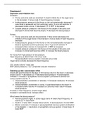
, Valsalva maneuver I
4 phases:
- Phase I: Pressure in the thorax on the vein/arteries/atria
increase but now stronger → blood is squeezed out from
pulmonary veins → go to left ventricle → increased
stroke volume → increased CO → MAP is increased
Heart rate decrease
- Phase IIa: Pressure inside the thorax is higher than pressure outside the
thorax (problem for blood vessels who have to transport blood from outside
the heart to the inside) → less blood is squeezed out from pulmonary veins →
go to left ventricle → decreased stroke volume → decreased CO → MAP is
decreased
Heart rate increase
- Phase IIb: recovery of MAP – recovery of heart rate
- Phase III: pressure in the thorax on the vein/arteries/atria decrease→ less
blood is squeezed out from pulmonary veins → go to left ventricle →
decreased stroke volume → decreased CO → MAP is decreased
Heart rate increase
- Phase IV: the venous inflow to the right ventricle is restored →
increased stroke volume → increased CO → MAP is increased
Heart rate decrease
When do you do a Valsalva → in labour, sometimes during sport
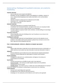
Blood pressure and posture
In lying position: blood pressure is equal in the body
In sitting position:
- Pressure in ankle is increased
- Pressure in arm stable around the same
- Pressure in ear is decreased
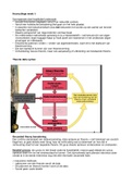
Pulse wave velocity
Forward (generated by contraction) and reflected (sum of lot of tiny
reflected wave generated by each branching point – long tail) wave
meet and interfere in arteries. Measured blood pressure is the sum of
these 2 pressure waves (green line)
Artery stiffen → pressure waves go back and forward quicker → come
together derived during systole → decreases in diastole lead to
decrease in filling of coronary arteries → increase risk of myocardial
ischemia
Peak of reflected wave is moved forward in time
In diastole the reflected pulse wave helps fill the coronary arteries
Expiration and inhalation test
Inhalation
• Thorax and atrial walls are stretched → stretch inhibits the of the vagal nerve
in the brainstem → sinus node → heart frequency increase
• Inside pressure: pressure in the thorax on the vein/arteries/atria decrease→
Less blood is squeezed out from pulmonary veins → go to left ventricle →
decreased stroke volume → decreased CO → MAP is decreased
• Outside pressure: pressure in the thorax on the outside of the aortic arch
decrease→ arterial wall becomes elastic → decrease the blood pressure
Exhale:
• Thorax and atrial walls are less stretched → less stretch decreases the
inhibition of the vagal nerve in the brainstem → sinus node → heart frequency
decrease
• Inside pressure: pressure in the thorax on the vein/arteries/atria increase→
blood is squeezed out from pulmonary veins → go to left ventricle →
increased stroke volume → increased CO → MAP is increased
• Outside pressure: pressure in the thorax on the outside of the aortic arch
increase → arterial wall becomes stiffer → increase the blood pressure
Air moves from high pressure to low pressure
Heart frequency is determined in the sinus node
Parasympathetic nervous system: innervate sinus node
Vagal nerve normally decrease the heart frequency
CO= stroke volume * heart rate
MAP (blood pressure) = CO * peripheral vascular resistance
Standing up test: baroreceptor reflex
Gravity will pool the blood in the legs → limit venous return to the heart → decrease
stroke volume → decreased CO → decrease blood pressure → baroreceptors
inhibited → increase in sympathetic nervous system (connected to arterial and
venous system) activity →
1. Vasoconstriction (constriction of arterials) → increased resistance → increase
blood pressure
2. Venoconstriction (constriction of veins) → push blood back to the heart →
increased stroke volume → increased CO (and thus heart rate)→ increase
blood pressure
Increase in heart frequency: chrome tropic effect
Increase in contractile force: inotropic effect
What lowers the blood pressure?
• Nocturnal period (night) → active vagus nerve → lowers heart frequency →
lowers CO → lower MAP
• Alcohol → lower ADH → lower blood volume → decreased CO → lower MAP
• Miction (urineren) → increase in parasympathetic nervous system (contraction
of the bladder) + inactivation of sympathetic nervous system in lower body →
lower MAP
, Valsalva maneuver I
4 phases:
- Phase I: Pressure in the thorax on the vein/arteries/atria
increase but now stronger → blood is squeezed out from
pulmonary veins → go to left ventricle → increased
stroke volume → increased CO → MAP is increased
Heart rate decrease
- Phase IIa: Pressure inside the thorax is higher than pressure outside the
thorax (problem for blood vessels who have to transport blood from outside
the heart to the inside) → less blood is squeezed out from pulmonary veins →
go to left ventricle → decreased stroke volume → decreased CO → MAP is
decreased
Heart rate increase
- Phase IIb: recovery of MAP – recovery of heart rate
- Phase III: pressure in the thorax on the vein/arteries/atria decrease→ less
blood is squeezed out from pulmonary veins → go to left ventricle →
decreased stroke volume → decreased CO → MAP is decreased
Heart rate increase
- Phase IV: the venous inflow to the right ventricle is restored →
increased stroke volume → increased CO → MAP is increased
Heart rate decrease
When do you do a Valsalva → in labour, sometimes during sport
Blood pressure and posture
In lying position: blood pressure is equal in the body
In sitting position:
- Pressure in ankle is increased
- Pressure in arm stable around the same
- Pressure in ear is decreased
Pulse wave velocity
Forward (generated by contraction) and reflected (sum of lot of tiny
reflected wave generated by each branching point – long tail) wave
meet and interfere in arteries. Measured blood pressure is the sum of
these 2 pressure waves (green line)
Artery stiffen → pressure waves go back and forward quicker → come
together derived during systole → decreases in diastole lead to
decrease in filling of coronary arteries → increase risk of myocardial
ischemia
Peak of reflected wave is moved forward in time
In diastole the reflected pulse wave helps fill the coronary arteries