NR 507 EDAPT HYPERSENSITIVITY GUIDE 2026 |
TYPES I–IV PATHOPHYSIOLOGY, CLINICAL CASES &
EXAM CORRECT QUSTIONS AND UPDATED SOLUTIONS
100% CORRECT
Week 1:Edapt Notes
1.
-Immediate hypersensitivity is mediated by IgE
antibodies, which resulting an allergy, anaphylaxis, or
atopic disease. The NP should expect the client to have a
type 1 hypersensitivity to recent medication use, which
can include these immediate reactions as clinical
manifestations: urticaria, wheezing, vomiting, and
diaphoresis.
2. are the primary effector cells and are responsible for
initiating and mediating
hypersensitivity reactions.
- Mast cells, type 1
characterized by the rapid release of
proinflammatory mediators like histamines,
,leukotrienes, and cytokines in response to allergen
exposure, mast cells are the primary effector cells
responsible for initiating and mediating type 1
hypersensitivity reactions.
3. hypersensitivity reactions involve the formation of
that can deposit in tissues, leading to complement
activation, inflammation, and tissue destruction.
-Type 3, immune complexes
-Type 3 hypersensitivity reactions involve the formation
of immune complexes that can deposit in tissues, leading
to complement activation and inflammation. This
process can cause tissue damage and is associated with
systemic lupus erythematosus (SLE) and serum sickness.
Type 1 reactions are mediated by IgE antibodies, and
type 2 are mediated by IgG or IgM antibodies. Type 4
reactions are activated by T- helper cells.
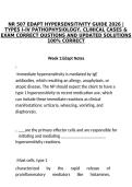
,Type 1 = Mediated by IgE antibodies
, Anaphylactic Reaction
1. Antigen
2. B-cell
3. Plasma cell
4. IgE
5. Mast cell
6. Histamine
A type 1 reaction is mediated by IgE
antibodies.
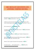
Type 2 = mediated by IgG or IgM antibodies
1. Anti-A antibodies in type B blood mix with type A blood
2. Antibodies attach to surface antigen of type A RBC
3. Complement activated; type A RBC cell wall attacked
4. Lysis of type A RBC
5. Phagocytosis
TYPES I–IV PATHOPHYSIOLOGY, CLINICAL CASES &
EXAM CORRECT QUSTIONS AND UPDATED SOLUTIONS
100% CORRECT
Week 1:Edapt Notes
1.
-Immediate hypersensitivity is mediated by IgE
antibodies, which resulting an allergy, anaphylaxis, or
atopic disease. The NP should expect the client to have a
type 1 hypersensitivity to recent medication use, which
can include these immediate reactions as clinical
manifestations: urticaria, wheezing, vomiting, and
diaphoresis.
2. are the primary effector cells and are responsible for
initiating and mediating
hypersensitivity reactions.
- Mast cells, type 1
characterized by the rapid release of
proinflammatory mediators like histamines,
,leukotrienes, and cytokines in response to allergen
exposure, mast cells are the primary effector cells
responsible for initiating and mediating type 1
hypersensitivity reactions.
3. hypersensitivity reactions involve the formation of
that can deposit in tissues, leading to complement
activation, inflammation, and tissue destruction.
-Type 3, immune complexes
-Type 3 hypersensitivity reactions involve the formation
of immune complexes that can deposit in tissues, leading
to complement activation and inflammation. This
process can cause tissue damage and is associated with
systemic lupus erythematosus (SLE) and serum sickness.
Type 1 reactions are mediated by IgE antibodies, and
type 2 are mediated by IgG or IgM antibodies. Type 4
reactions are activated by T- helper cells.
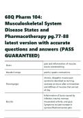
,Type 1 = Mediated by IgE antibodies
, Anaphylactic Reaction
1. Antigen
2. B-cell
3. Plasma cell
4. IgE
5. Mast cell
6. Histamine
A type 1 reaction is mediated by IgE
antibodies.
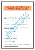
Type 2 = mediated by IgG or IgM antibodies
1. Anti-A antibodies in type B blood mix with type A blood
2. Antibodies attach to surface antigen of type A RBC
3. Complement activated; type A RBC cell wall attacked
4. Lysis of type A RBC
5. Phagocytosis