BMS4004 Introduction to Physiology 2019-20
Lab Report
Please complete all questions. The questions are worth a total of 30 marks.
Neuro-muscular physiology
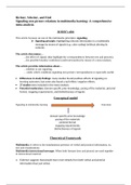
1. Using a figure explain how the myotatic reflex is activated by hitting the Achilles tendon
and describe the role of each element in the figure.
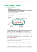
The myotatic reflex, also referred to as the knee-jerk, is a response to a stimulus that does
not require the involvement of consciousness and is a response to the changes in the length
of the muscle. This means that the response is involuntary. This occurs due to a stimulus,
hitting the Achilles tendon which is situated below the knee-cap. The first stage of this reflex
is the afferent stage where the afferent nerves sends information of the stimulus up to the
central nervous system where receptors receive this information. When a force stretches
the muscle, the muscle intrafusal muscle fibres can alter the length of the spindle by γ
motor efferents, to keep the stretch sensitive area under tension. This increases the firing
rate of the 1a afferent. The second stage of the reflex is the efferent stage. The efferent
nerve carrier information away from the central nervous system which causes a response in
the periphery as seen in figure 1. This increased 1a afferent activity in the spinal cord
causing increased activity of the motor neurons in that muscle. This contracts the muscle
back to its original length. This response is your leg kicking out. This reflex occurs on the
Fig 1: Myotatic Reflex
, same side of the body and the brain is not a factor within the chain of events.
(3 marks)
2. Describe how the EMG signal (a voltage) arises from muscle activity.
An action potential causes the voltage gated calcium channels to open Ca2+ release into
the presynaptic terminal and causes the myosin-actin cross-bridge to cycle. Vesicles that
are filled with acetylcholine fuse with the cell membrane. This releases the ACh in the
synaptic cleft Na+ currents underly the upstroke of the action potential which causes a
brief extracellular negative potential. The ACh then binds to the nicotinic acetylcholine
receptors on the endplate. This opens cation channels that cause a depolarisation EPP. If
this EPP exceeds the action potential threshold, an action potential is generated. This
spreads across the muscle cell membrane and propagates into the system called the T-
tubule. This causes a release of Ca2+ from the sarcoplasmic reticulum. When this occurs,
the CA2+ channels connect to a voltage sensor on the T-tubule. The binding of Ca2+ to
troponin moves tropomyosin. A new actin binding site on the actin strand is revealed.
The myosin head bends, and this process is assisted by the ATP binding to the actin
strand. It bends backwards to travel along the actin strand. The sarcomere of the
microfibril shortens with the myocyte. All myocytes using the same motor neurons will
contract in unison. Multiple motor units together cause a compound muscle potential.
An EMG translates these signals into a graph or numbers to allow for analysis.
(2 marks)
Lab Report
Please complete all questions. The questions are worth a total of 30 marks.
Neuro-muscular physiology
1. Using a figure explain how the myotatic reflex is activated by hitting the Achilles tendon
and describe the role of each element in the figure.
The myotatic reflex, also referred to as the knee-jerk, is a response to a stimulus that does
not require the involvement of consciousness and is a response to the changes in the length
of the muscle. This means that the response is involuntary. This occurs due to a stimulus,
hitting the Achilles tendon which is situated below the knee-cap. The first stage of this reflex
is the afferent stage where the afferent nerves sends information of the stimulus up to the
central nervous system where receptors receive this information. When a force stretches
the muscle, the muscle intrafusal muscle fibres can alter the length of the spindle by γ
motor efferents, to keep the stretch sensitive area under tension. This increases the firing
rate of the 1a afferent. The second stage of the reflex is the efferent stage. The efferent
nerve carrier information away from the central nervous system which causes a response in
the periphery as seen in figure 1. This increased 1a afferent activity in the spinal cord
causing increased activity of the motor neurons in that muscle. This contracts the muscle
back to its original length. This response is your leg kicking out. This reflex occurs on the
Fig 1: Myotatic Reflex
, same side of the body and the brain is not a factor within the chain of events.
(3 marks)
2. Describe how the EMG signal (a voltage) arises from muscle activity.
An action potential causes the voltage gated calcium channels to open Ca2+ release into
the presynaptic terminal and causes the myosin-actin cross-bridge to cycle. Vesicles that
are filled with acetylcholine fuse with the cell membrane. This releases the ACh in the
synaptic cleft Na+ currents underly the upstroke of the action potential which causes a
brief extracellular negative potential. The ACh then binds to the nicotinic acetylcholine
receptors on the endplate. This opens cation channels that cause a depolarisation EPP. If
this EPP exceeds the action potential threshold, an action potential is generated. This
spreads across the muscle cell membrane and propagates into the system called the T-
tubule. This causes a release of Ca2+ from the sarcoplasmic reticulum. When this occurs,
the CA2+ channels connect to a voltage sensor on the T-tubule. The binding of Ca2+ to
troponin moves tropomyosin. A new actin binding site on the actin strand is revealed.
The myosin head bends, and this process is assisted by the ATP binding to the actin
strand. It bends backwards to travel along the actin strand. The sarcomere of the
microfibril shortens with the myocyte. All myocytes using the same motor neurons will
contract in unison. Multiple motor units together cause a compound muscle potential.
An EMG translates these signals into a graph or numbers to allow for analysis.
(2 marks)