Science Medicine Cardiology Save
SAEM EXAM QUESTIONS 2024-2025 ACTUAL EXAM 400
REAL EXAM QUESTIONS AND CORRECT DETAILED
ANSWERS WITH RATIONALES (VERIFIED ANSWERS) ,
SAEM Practice 2020, Edited SAEM Exam Set
Leave the first rating
Students also studied
30 terms 77 terms 56 terms Teacher
Preview Preview Preview
Terms in this set (524)
"Which coronary vessel is usually the cause of the "A. left anterior descending
(LAD) myocardial infarction in a patient with ST elevation in V1,
V2, and V3? The answer is A. This EKG pattern is consistent with that of anterior wall
A. left anterior descending (LAD) myocardial infarction (MI). The LAD supplies the anterior wall of the myocardium.
B.left circumflex artery The left circumflex artery, the LAD, or a branch of the RCA supplies the lateral wall
C. posterior descending branch of the right coronary of the left ventricle. Proximal occlusion of the LAD will give ST elevation in leads
artery V1-6, aVL and I (an anterolateral MI). Occlusion of a branch of the RCA will result
D.right coronary artery (RCA) in an inferolateral MI (ST elevation in leads II, III, aVF and I, aVL, V5 and V6). The
E.right ventricular branch of the right coronary artery" RCA supplies the inferior wall and SA node. Occlusion in leads II, III and aVF
causes an inferior MI. The right ventricle is usually supplied by the RCA or, less
commonly, a dominant left circumflex. ST elevation in leads V4 and V5 of a
right- side leads EKG suggests infarction of the right ventricle. A posterior MI
(ST depression in V1-V3) results from occlusion of the RCA, its posterior
descending branch, or a dominant left circumflex."
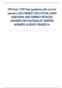
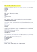
"A 51-year-old male with long-standing hypertension "C. CT of the chest with IV contrast
presents with abrupt onset of severe chest pain radiating
to the back. He describes a tearing sensation. Vital signs "CT of the chest is the test most often used to confirm the diagnosis of
aortic are HR 110, BP 175/105, RR 20, T 37.4. EKG shows LVH. dissection. CT is readily available in most Emergency
Departments, and has a CBC, electrolytes, BUN/Creatinine are all normal. CXR is sensitivity of 83-98% and specificity of 87-100% for
aortic dissection (highest
as shown below. What diagnostic test would be most accuracy with helical scans). Other benefits associated with the use of CT
include appropriate for making a definitive diagnosis at this time? the ability to identify intramural thrombus, pericardial effusion,
and potentially [image shows CXR w/ wide mediastinum] reveal another etiology for the patient's pain. The major disadvantage of CT is
the
need for iodinated contrast, which requires normal renal function.""
A. MRI of the thoracic spine
B.Aortogram
To get the actual and other Exams contact ()
, C. CT of the chest with IV contrast
D.Esophagram using Gastrograffin"
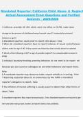
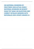
"A 60 year old male presented to the emergency "B. ventricular
tachycardia department with chest pain. He subsequently became
unresponsive. The monitor shows the rhythm below. The The answer is B. Ventricular tachycardia is wide and complex. It is
distinguished rhythm is: from supraventricular tachycardia by width and morphology of the QRS
[image monomorphic wide QRS tachycardia with no p complexes. (Though there are numerous exceptions, supraventricular tachycardias
waves] usually exhibit narrow QRS complexes with morphology similar to that when the
A. sinus tachycardia patient is in sinus rhythm.)"
B.ventricular tachycardia
C. atrial fibrillation with rapid ventricular response
D.atrial flutter"
"A 64 year old female presents to the emergency "A. hypertensive crisis
department with chief complaints of occipital headache
and chest pain. Physical examination reveals a blood The answer is A. Elevated blood pressure in the setting of optic disk edema
is a pressure of 200/118 as well as edema of the optic disk. Of hallmark of malignant hypertension (also known as hypertensive
emergency or the diagnoses below, the most likely is: hypertensive crisis). While hypertensive urgency is not consistently defined in
the
A. hypertensive crisis medical literature, this patient's presentation indicates that there is some end-
B.acute hypertensive (non-emergency/non-urgency) organ damage and thus the diagnosis is malignant hypertension. The white-coat""
episode syndrome, in which patients' blood pressures are elevated only in the
clinical
C. hypertensive urgency setting and not at home, has been shown to account for as many as a fifth of all
D.moderate hypertension cases of newly diagnosed ""hypertension."" Understanding of this phenomenom is
E.white-coat hypertension" important for emergency physicians, since its frequency explains why
patients should not be given a diagnosis of new-onset hypertension
based on E.D. measurements."""
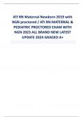
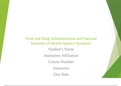
"A 14 year old presents just after smoking crack cocaine "D.
Pneumomediastinum and complains of chest pain. He describes it as sharp and
stabbing in the middle of his chest. His EKG is normal. The The answer is D. Look closely along the right heart border and mediastinum.
There intern reads the CXR as "negative" but your supervising is a thin strip of air. Pneumomediastinum and pneumopericardium
result from resident asks you to have another look (see Figure), after Valsalva maneuvers, barotrauma, asthma, and cocaine inhalation
from positive which you make the diagnosis of: pressure devices. On physical exam there may be a Hamman's sign or
mediastinal [image: big round heart, black in mediastinum, widened] crunch heard over the precordium. Westermark's sign is dilation of
pulmonary photo courtesy of eMedicine.com vessels proximal to a pulmonary embolism resulting in a cut-off appearance
of
A. Pneumonia the vessel on CXR."
B.Aortic dissection
C. Congestive heart failure
D.Pneumomediastinum"
To get the actual and other Exams contact ()
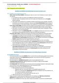
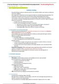
, "A 22 year old presents with chest pain and the following "A.
Cocaine EKG:
The answer is A. Cocaine toxicity can cause a variety of cardiovascular sequelae
[image: Septal ST elevations] including: cardiac dysrhythmias, coronary artery vasospasm, myocardial
ischemia/infarction, and aortic dissection. The central nervous system is also
He reports no past medical history and no family history commonly involved with seizures, intracranial hemorrhages/infarctions and
of medical problems. Which substance should you hypertensive encephalopathy being common. Mesenteric ischemia can occur as
specifically question him about using? well
as rhabdomyolysis."
A. Cocaine
B.Heroin
C. Methamphetamine
D.Ecstasy"
To get the actual and other Exams contact ()
SAEM EXAM QUESTIONS 2024-2025 ACTUAL EXAM 400
REAL EXAM QUESTIONS AND CORRECT DETAILED
ANSWERS WITH RATIONALES (VERIFIED ANSWERS) ,
SAEM Practice 2020, Edited SAEM Exam Set
Leave the first rating
Students also studied
30 terms 77 terms 56 terms Teacher
Preview Preview Preview
Terms in this set (524)
"Which coronary vessel is usually the cause of the "A. left anterior descending
(LAD) myocardial infarction in a patient with ST elevation in V1,
V2, and V3? The answer is A. This EKG pattern is consistent with that of anterior wall
A. left anterior descending (LAD) myocardial infarction (MI). The LAD supplies the anterior wall of the myocardium.
B.left circumflex artery The left circumflex artery, the LAD, or a branch of the RCA supplies the lateral wall
C. posterior descending branch of the right coronary of the left ventricle. Proximal occlusion of the LAD will give ST elevation in leads
artery V1-6, aVL and I (an anterolateral MI). Occlusion of a branch of the RCA will result
D.right coronary artery (RCA) in an inferolateral MI (ST elevation in leads II, III, aVF and I, aVL, V5 and V6). The
E.right ventricular branch of the right coronary artery" RCA supplies the inferior wall and SA node. Occlusion in leads II, III and aVF
causes an inferior MI. The right ventricle is usually supplied by the RCA or, less
commonly, a dominant left circumflex. ST elevation in leads V4 and V5 of a
right- side leads EKG suggests infarction of the right ventricle. A posterior MI
(ST depression in V1-V3) results from occlusion of the RCA, its posterior
descending branch, or a dominant left circumflex."
"A 51-year-old male with long-standing hypertension "C. CT of the chest with IV contrast
presents with abrupt onset of severe chest pain radiating
to the back. He describes a tearing sensation. Vital signs "CT of the chest is the test most often used to confirm the diagnosis of
aortic are HR 110, BP 175/105, RR 20, T 37.4. EKG shows LVH. dissection. CT is readily available in most Emergency
Departments, and has a CBC, electrolytes, BUN/Creatinine are all normal. CXR is sensitivity of 83-98% and specificity of 87-100% for
aortic dissection (highest
as shown below. What diagnostic test would be most accuracy with helical scans). Other benefits associated with the use of CT
include appropriate for making a definitive diagnosis at this time? the ability to identify intramural thrombus, pericardial effusion,
and potentially [image shows CXR w/ wide mediastinum] reveal another etiology for the patient's pain. The major disadvantage of CT is
the
need for iodinated contrast, which requires normal renal function.""
A. MRI of the thoracic spine
B.Aortogram
To get the actual and other Exams contact ()
, C. CT of the chest with IV contrast
D.Esophagram using Gastrograffin"
"A 60 year old male presented to the emergency "B. ventricular
tachycardia department with chest pain. He subsequently became
unresponsive. The monitor shows the rhythm below. The The answer is B. Ventricular tachycardia is wide and complex. It is
distinguished rhythm is: from supraventricular tachycardia by width and morphology of the QRS
[image monomorphic wide QRS tachycardia with no p complexes. (Though there are numerous exceptions, supraventricular tachycardias
waves] usually exhibit narrow QRS complexes with morphology similar to that when the
A. sinus tachycardia patient is in sinus rhythm.)"
B.ventricular tachycardia
C. atrial fibrillation with rapid ventricular response
D.atrial flutter"
"A 64 year old female presents to the emergency "A. hypertensive crisis
department with chief complaints of occipital headache
and chest pain. Physical examination reveals a blood The answer is A. Elevated blood pressure in the setting of optic disk edema
is a pressure of 200/118 as well as edema of the optic disk. Of hallmark of malignant hypertension (also known as hypertensive
emergency or the diagnoses below, the most likely is: hypertensive crisis). While hypertensive urgency is not consistently defined in
the
A. hypertensive crisis medical literature, this patient's presentation indicates that there is some end-
B.acute hypertensive (non-emergency/non-urgency) organ damage and thus the diagnosis is malignant hypertension. The white-coat""
episode syndrome, in which patients' blood pressures are elevated only in the
clinical
C. hypertensive urgency setting and not at home, has been shown to account for as many as a fifth of all
D.moderate hypertension cases of newly diagnosed ""hypertension."" Understanding of this phenomenom is
E.white-coat hypertension" important for emergency physicians, since its frequency explains why
patients should not be given a diagnosis of new-onset hypertension
based on E.D. measurements."""
"A 14 year old presents just after smoking crack cocaine "D.
Pneumomediastinum and complains of chest pain. He describes it as sharp and
stabbing in the middle of his chest. His EKG is normal. The The answer is D. Look closely along the right heart border and mediastinum.
There intern reads the CXR as "negative" but your supervising is a thin strip of air. Pneumomediastinum and pneumopericardium
result from resident asks you to have another look (see Figure), after Valsalva maneuvers, barotrauma, asthma, and cocaine inhalation
from positive which you make the diagnosis of: pressure devices. On physical exam there may be a Hamman's sign or
mediastinal [image: big round heart, black in mediastinum, widened] crunch heard over the precordium. Westermark's sign is dilation of
pulmonary photo courtesy of eMedicine.com vessels proximal to a pulmonary embolism resulting in a cut-off appearance
of
A. Pneumonia the vessel on CXR."
B.Aortic dissection
C. Congestive heart failure
D.Pneumomediastinum"
To get the actual and other Exams contact ()
, "A 22 year old presents with chest pain and the following "A.
Cocaine EKG:
The answer is A. Cocaine toxicity can cause a variety of cardiovascular sequelae
[image: Septal ST elevations] including: cardiac dysrhythmias, coronary artery vasospasm, myocardial
ischemia/infarction, and aortic dissection. The central nervous system is also
He reports no past medical history and no family history commonly involved with seizures, intracranial hemorrhages/infarctions and
of medical problems. Which substance should you hypertensive encephalopathy being common. Mesenteric ischemia can occur as
specifically question him about using? well
as rhabdomyolysis."
A. Cocaine
B.Heroin
C. Methamphetamine
D.Ecstasy"
To get the actual and other Exams contact ()