THE BASIS OF NEURAL SIGNALS
In the scientific literature, scholars attacked the usefulness of brain imaging. They argue that brain
imaging only informs us about where mental functions are in the brain. The terms “neophrenology” are
often used in this context, referring to the phrenologists of the nineteenth century claiming that outer
features of the skull were related to mental functions. Phrenology was a pseudoscience because the claim
was never proved empirically, so comparison is not fair. Still, it is valid to ask if knowing where things
are is relevant for, e.g., psychology and cognitive science:
▪ Highly relevant as a first step, because we need to know where a mental function resides in the
brain before we can study it further through neuroscientific techniques.
▪ The next step is to investigate how the mental function is implemented through neural networks
and circuitry:
= Relevant for constraining psychological/cognitive models. Contrary to what is suggested by
denoting brain imaging as neo-phrenology, brain scans are not limited to localization in the narrow
sense and can also help in this next step, often together with other neuroscientific methods.
INFORMATION TRANSFER IN NEURONS
There are many kinds of neurons that typically have the following parts:
▪ Dendritic tree
▪ Soma (cell body)
▪ Axon
Grey matter
The brain is organized in such a way that the cell bodies of neurons are concentrated in particular
structures. These structures look grayish in the living brain, is called gray matter. The cerebral cortex is a
sheet of gray matter and thus contains cell bodies. Other concentrations of cell bodies beneath the cortex,
subcortical structures, are called nuclei.
White matter
Some neurons have short axons that remain in the gray matter, but many connect to distant neurons
through long axons. All these long axons together make up the white matter. Underneath the cortex, this
white matter takes up a large volume; in the more peripheral nervous system, the axons form nerve
bundles and tracts.
COMMUNICATION BETWEEN NEURONS
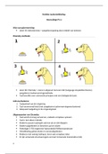
Figure shows at the right a schematic neuron in red with the same major components. This neuron
receives input from other neurons, a few of which are shown on the left.
The neurons in red are neurons that provide excitatory
signals that make the receiving neuron become more
“active.”
The neuron in blue represents an inhibitory neuron that
makes the receiving neuron less active.
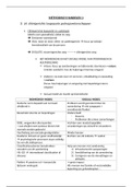
Resting state
Without any input, our neuron is at rest.
This resting state is characterized by a
resting potential at the cell membrane. This
resting potential is an electrical potential
difference between the inside and the outside of the neuron. At rest, this potential
difference is -70 millivolts (mV).
1
, This resting state is the starting point of the graph.
Input from other neurons
Neurotransmitter
Our neuron receives input from other neurons by the delivery of a chemical substance referred to as a
neurotransmitter at the synapses (contact points between neurons) in the dendritic tree of our neuron.
Receptor
Receptors in the membrane of our neuron react to these neurotransmitters and disturb the resting
potential. The direction of this effect differs between neurotransmitters and, depending on which
neurotransmitter is released, neurons are categorized as excitatory or inhibitory.
Depolarization
The neurotransmitter input from an excitatory neuron will make the potential difference less
negative, so the -70 mV might become -65 mV. In the cerebral cortex, glutamate is an excitatory
neurotransmitter.
Hyperpolarization
The neurotransmitter from an inhibitory neuron will make the potential difference more
negative. In the cerebral cortex, a molecule known as GABA is the most prominent inhibitory
neurotransmitter.
Axon hillock
The changes in the potential difference originate in the dendritic tree of our neuron, but are
transmitted throughout the cell membrane of the soma, toward the point where the axon begins. This
point is called the “axon hillock.”
The action potential
A sequence of events occurs at the cell membrane when the potential difference reaches a critical level,
typically at -55 mV: When the difference between the inside and the outside of the neuron
becomes this small:
Results in a sudden further decrease of the potential difference, an overshoot so that the
difference becomes positive,
Then there is a very quick restoration of a negative difference. These rapid changes in the potential
take a very characteristic form, known as the action potential (AP). Given their sharpness, APs are
sometimes also called “spikes.”
Figure shows how an action potential works:
The rest potential difference is -70 mV. This is the starting point in the
schematic potential function
The neurotransmitter input from an excitatory neuron will make the potential
difference less negative, depolarization, so the -70 mV might become -
65 mV.
The neurotransmitter from an inhibitory neuron will have the opposite effect
and make the potential difference more negative, hyperpolarization.
The potential difference reaches a critical level, typically at -55 mV
sequence of events occurs at the cell membrane
The schematic potential below shows three such
action potentials: The figure shows three excitatory
neurons in red and one inhibitory neuron in blue.
2
In the scientific literature, scholars attacked the usefulness of brain imaging. They argue that brain
imaging only informs us about where mental functions are in the brain. The terms “neophrenology” are
often used in this context, referring to the phrenologists of the nineteenth century claiming that outer
features of the skull were related to mental functions. Phrenology was a pseudoscience because the claim
was never proved empirically, so comparison is not fair. Still, it is valid to ask if knowing where things
are is relevant for, e.g., psychology and cognitive science:
▪ Highly relevant as a first step, because we need to know where a mental function resides in the
brain before we can study it further through neuroscientific techniques.
▪ The next step is to investigate how the mental function is implemented through neural networks
and circuitry:
= Relevant for constraining psychological/cognitive models. Contrary to what is suggested by
denoting brain imaging as neo-phrenology, brain scans are not limited to localization in the narrow
sense and can also help in this next step, often together with other neuroscientific methods.
INFORMATION TRANSFER IN NEURONS
There are many kinds of neurons that typically have the following parts:
▪ Dendritic tree
▪ Soma (cell body)
▪ Axon
Grey matter
The brain is organized in such a way that the cell bodies of neurons are concentrated in particular
structures. These structures look grayish in the living brain, is called gray matter. The cerebral cortex is a
sheet of gray matter and thus contains cell bodies. Other concentrations of cell bodies beneath the cortex,
subcortical structures, are called nuclei.
White matter
Some neurons have short axons that remain in the gray matter, but many connect to distant neurons
through long axons. All these long axons together make up the white matter. Underneath the cortex, this
white matter takes up a large volume; in the more peripheral nervous system, the axons form nerve
bundles and tracts.
COMMUNICATION BETWEEN NEURONS
Figure shows at the right a schematic neuron in red with the same major components. This neuron
receives input from other neurons, a few of which are shown on the left.
The neurons in red are neurons that provide excitatory
signals that make the receiving neuron become more
“active.”
The neuron in blue represents an inhibitory neuron that
makes the receiving neuron less active.
Resting state
Without any input, our neuron is at rest.
This resting state is characterized by a
resting potential at the cell membrane. This
resting potential is an electrical potential
difference between the inside and the outside of the neuron. At rest, this potential
difference is -70 millivolts (mV).
1
, This resting state is the starting point of the graph.
Input from other neurons
Neurotransmitter
Our neuron receives input from other neurons by the delivery of a chemical substance referred to as a
neurotransmitter at the synapses (contact points between neurons) in the dendritic tree of our neuron.
Receptor
Receptors in the membrane of our neuron react to these neurotransmitters and disturb the resting
potential. The direction of this effect differs between neurotransmitters and, depending on which
neurotransmitter is released, neurons are categorized as excitatory or inhibitory.
Depolarization
The neurotransmitter input from an excitatory neuron will make the potential difference less
negative, so the -70 mV might become -65 mV. In the cerebral cortex, glutamate is an excitatory
neurotransmitter.
Hyperpolarization
The neurotransmitter from an inhibitory neuron will make the potential difference more
negative. In the cerebral cortex, a molecule known as GABA is the most prominent inhibitory
neurotransmitter.
Axon hillock
The changes in the potential difference originate in the dendritic tree of our neuron, but are
transmitted throughout the cell membrane of the soma, toward the point where the axon begins. This
point is called the “axon hillock.”
The action potential
A sequence of events occurs at the cell membrane when the potential difference reaches a critical level,
typically at -55 mV: When the difference between the inside and the outside of the neuron
becomes this small:
Results in a sudden further decrease of the potential difference, an overshoot so that the
difference becomes positive,
Then there is a very quick restoration of a negative difference. These rapid changes in the potential
take a very characteristic form, known as the action potential (AP). Given their sharpness, APs are
sometimes also called “spikes.”
Figure shows how an action potential works:
The rest potential difference is -70 mV. This is the starting point in the
schematic potential function
The neurotransmitter input from an excitatory neuron will make the potential
difference less negative, depolarization, so the -70 mV might become -
65 mV.
The neurotransmitter from an inhibitory neuron will have the opposite effect
and make the potential difference more negative, hyperpolarization.
The potential difference reaches a critical level, typically at -55 mV
sequence of events occurs at the cell membrane
The schematic potential below shows three such
action potentials: The figure shows three excitatory
neurons in red and one inhibitory neuron in blue.
2