OPTHALMOLOGY
EYE ANATOMY ............................................................................................................................. 2
LIGHT PATHWAY ........................................................................................................................ 10
PATHOLOGIES ............................................................................................................................ 11
PHARMACOKINETICS ................................................................................................................. 24
PHARMACODYNAMICS .............................................................................................................. 26
MEDICATION ............................................................................................................................. 27
1
, EYE ANATOMY
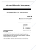
EYEBALL
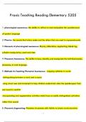
- Eyeball is made of 3 layers (picture)
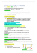
ORBIT Roof
• Frontal
• Lesser wing of sphenoid
Floor
• Maxilla
• Palatine
• Zygomatic
Medial
• Ethmoid
• Maxilla
• Sphenoid
Lateral
• Zygomatic
• Greater wing of sphenoid
- The medical wall is the thinnest, followed by the floor of the mouth which is
strengthened by the ethmoid sinuses.
- Floor is most vulnerable to fractures when there is direct force on the ocular globe.
This is because it is thin and unsupported
2
, - All the orbital walls are CURVILINEAR in shape. This allows protection of eyeball and
cushion it vs blunt force.
- When there is a blowout fracture: incarceration of rectus muscles, Oedema
ecchymosis, orbital compartment syndrome, upgazed restriction.
SCLERA - It is where bilirubin accumulates (especially the dense connective tissue)
- Divided in:
o Episclera (dense CT)
o Sclera propria (collagen)
o Lamina fusca (pigmented)
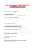
EYELIDS F(x): offer protection and distribute tear film.
Anatomy:
- Skin
- Orbicularis oculi (CN VII)
o Eyelid closure, tear regulation
(squeeze, empty)
- Submuscular adipose tissue (SMFAT)
- Orbital septum
o Divides orbital content from lid
content
o Contains the spread of infection
- Tarsal plates (connective tissue)
o Inferior & superior – act as a scaffold
o Meibomian glands
- Levator apparatus (CN III)
o Levator Palpebrae Superioris (Skeletal)
o Superior Tarsal Muscle (Muller’s muscle, SNS)
o Inferior Tarsal Muscle (Muller’s muscle, SNS)
- Conjunctiva
PTOSIS:
- Complete: paralysis of LPS due to CN3 lesion (somatic nerves and skeletal muscles)
- Partial: Paralysis of Muller’s Muscle (in the tarsal plate) due to Horner’s
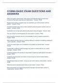
LACRIMAL - Contribute to aqueous layer of the tear film
SYSTEM - Lipid layer – superficial, oily (MGs)
•
- Aqueous layer – substrates, immune (lacrimal)
- Mucinous layer – adhesion (epithelium)
Lacrimal gland → Punctum → Canaliculus → Common canaliculus
→ lacrimal sac → nasolacrimal duct
CORNEA - 5 layers and contributes to 80% refraction
3
EYE ANATOMY ............................................................................................................................. 2
LIGHT PATHWAY ........................................................................................................................ 10
PATHOLOGIES ............................................................................................................................ 11
PHARMACOKINETICS ................................................................................................................. 24
PHARMACODYNAMICS .............................................................................................................. 26
MEDICATION ............................................................................................................................. 27
1
, EYE ANATOMY
EYEBALL
- Eyeball is made of 3 layers (picture)
ORBIT Roof
• Frontal
• Lesser wing of sphenoid
Floor
• Maxilla
• Palatine
• Zygomatic
Medial
• Ethmoid
• Maxilla
• Sphenoid
Lateral
• Zygomatic
• Greater wing of sphenoid
- The medical wall is the thinnest, followed by the floor of the mouth which is
strengthened by the ethmoid sinuses.
- Floor is most vulnerable to fractures when there is direct force on the ocular globe.
This is because it is thin and unsupported
2
, - All the orbital walls are CURVILINEAR in shape. This allows protection of eyeball and
cushion it vs blunt force.
- When there is a blowout fracture: incarceration of rectus muscles, Oedema
ecchymosis, orbital compartment syndrome, upgazed restriction.
SCLERA - It is where bilirubin accumulates (especially the dense connective tissue)
- Divided in:
o Episclera (dense CT)
o Sclera propria (collagen)
o Lamina fusca (pigmented)
EYELIDS F(x): offer protection and distribute tear film.
Anatomy:
- Skin
- Orbicularis oculi (CN VII)
o Eyelid closure, tear regulation
(squeeze, empty)
- Submuscular adipose tissue (SMFAT)
- Orbital septum
o Divides orbital content from lid
content
o Contains the spread of infection
- Tarsal plates (connective tissue)
o Inferior & superior – act as a scaffold
o Meibomian glands
- Levator apparatus (CN III)
o Levator Palpebrae Superioris (Skeletal)
o Superior Tarsal Muscle (Muller’s muscle, SNS)
o Inferior Tarsal Muscle (Muller’s muscle, SNS)
- Conjunctiva
PTOSIS:
- Complete: paralysis of LPS due to CN3 lesion (somatic nerves and skeletal muscles)
- Partial: Paralysis of Muller’s Muscle (in the tarsal plate) due to Horner’s
LACRIMAL - Contribute to aqueous layer of the tear film
SYSTEM - Lipid layer – superficial, oily (MGs)
•
- Aqueous layer – substrates, immune (lacrimal)
- Mucinous layer – adhesion (epithelium)
Lacrimal gland → Punctum → Canaliculus → Common canaliculus
→ lacrimal sac → nasolacrimal duct
CORNEA - 5 layers and contributes to 80% refraction
3