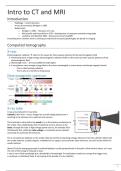
Intro to CT and MRI
Introduction
- Radiology = recent discovery
- X-rays discovered by Röntgen in 1895
- Nobel prizes
o Röntgen in 1901 – discovery of x-rays
o McCormack and Hounsfield in 1979 – development of computer assisted tomography
o Lauterbur and Mansfiel 2003 – discoveries concerning MRI
Everything from a broken wrist to viewing at anatomical structures/pathologies are based on imaging
Computed tomography
X-rays
Electromagnetic radiation → refers to the waves (or their quanta, photons) of the electromagnetic field
• Penetrating form of high-energy electromagnetic radiation Refers to the waves (or their quanta, photons) of the
electromagnetic field
• Wavelength: 0,01 – 10 nm (1/10000 of visible light)
• X-ray photons carry enough energy (due to the short wavelength) to ionize atoms and disrupt magnetic bonds
- This is called ionizing radiation
- That is why it is harmful to living tissue
Electromagnetic spectrum
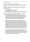
X-ray tube
X-rays are produced in X-ray tubes.
Cathode (=thin wire – red on image) has a small voltage placed over it
resulting in the emission of an electron into vacuum.
This electrode is attracted to the anode (+), so the electron accelerates to
the anode, thus establishing a flow of electrical current, known as the
beam, through the tube. A high voltage power source, for example 30 to
150 kilovolts (kV), called the tube voltage, is connected across cathode
and anode to accelerate the electrons.
The X-ray spectrum depends on the anode material and the accelerating voltage. Electrons from the cathode collide with
the anode material, usually tungsten, molybdenum or copper, and accelerate other electrons, ions and nuclei within the
anode material.
About 1% of the energy generated is emitted/radiated, usually perpendicular to the path of the electron beam, as X-rays.
The rest of the energy is released as heat.
x-rays is not a fixed wavelength but a bandwidth of wavelength → small patient needs less energy than bigger ones
x-ray beam is collimated/ fixed → narrowing of the bundle of x-ray radiation
1
,CT-scan
Same principle as X-ray but with CT scan you make many images from different
orientations
Inside: x-ray tube and a detector
Patient is in the middle
Gantric → the thing that moves around the patient
The images you get are multiple fine slices of your structure and you can look at it
from different aspects
Image reconstruction
How do you get a cross-sectional image from data acquired in different directions?
Back projection: images acquired in each direction are smeared out over the image we are going to reconstruct
Example reconstruction of dot
o 2 projections: a cross with a brighter square in the middle
o 4 or 8 projections: looks more like a circle but has a star artefact
o 16 or 64 projections: looks like the real image, but still some star artefact
In real CT more than 500 projections are used → Result = blurry image because you
smear out the signal along the axis so there is signal from the different directions
Difference with filtered back projection: for each image gets a sharpening filter
Filtered back projection: → the filtering Is done before the images are
produced
• High pass/sharpening filters are applied before back projection
• Picks up sharp edges in the projection, ignores flat areas (areas without
contrast)
Reconstruction kernel – different filters for different images:
• Sharp filter used for lungs and bones
• Soft filter (kernel) used for brain and abdomen
2
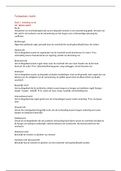
,Iterative reconstruction
The CT scans measures the total signals (12, 8, 7) = sums of the matrix. To
reconstruct the original density values (light grey – 5,7,…) of the matrix the
following needs to be done:
1. Grey = unknowns → original data of 4 pixels
2. The measured signals are split between the two boxes
→ eg: vertical projections – the sum is 11 and 9 so these are split over
the boxes into 2x 5,5 and 2x 4,5 (which are of course not the real
values)
3. Then we look at the horizontal projections
a. Top row: if we sum 4,5 and 5,5 we get 10, but we measured 12 ,
so 2 more need to be added. These are divided over both boxes,
so in each box +1 → 4,5 becomes 5,5 and 5,5 becomes 6,5
b. Bottom row: the sum of the boxes is 10, but we measures 8 so -2
needs to be applied → 5,5 becomes 4,5 and 4,5 becomes 3,5
4. Then we look at the diagonals
a. From bottom left to top right: sum is 10, but we measured 13, so 3 needs to be added → 5,5 becomes 7 and
4,5 becomes 6
b. From bottom right to top left: sum = 10 but we measured 7 --> -3 --> 6,5 becomes 5 and 3,5 becomes 2
Now we have the correct values, so after a few iterations you come to the correct solution. Of course, if you have a larger
matrix it becomes more complicated. The more accurate the data (the more measurements done and the more radiation
used), the more accurate the solution.
Advantage: comes to a better solution than filtered back →
Lower radiation dose can be used (less x-rays)
Conclusion:
1. You start off with filtered back projection = 1 guess
2. Foward projection: calculates – if this would be the right result what
would you measure (compare to measured data)
3. Back projection
4. Repeat the iterations
Computed tomography
Acquisition
Density = x-ray radiation passing though the tissue
• High attenuation (absorption) = bright pixel
- Metal
- Bone (not as white as metal)
• Low attenuation = dark pixel
- Air
- Fat (not as dark as air-
• Brain is somewhere in between
White = less x-rays
Dark = more x-rays
3
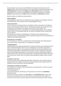
, Analysis
Hounsfield units: a quantitative scale for describing radiodensity
used in CT
• Water = 0
• Air = -1000
• All the other grey values are somewhere in between, so
these can differ between patients
CT data sets have a very high dynamic range which must be
reduced for display or printing. This is typically done via a process
of "windowing", which maps a range ("window") of pixel values to a grayscale ramp.
For example, CT images of the brain are commonly viewed with a window extending from 0 HU to 80 HU. Pixel
values of 0 and lower, are displayed as black; values of 80 and higher are displayed as white; values within the
window are displayed as a grey intensity proportional to position within the window
The window used for display must be matched to the X-ray density of the object of interest, in order to optimize
the visible detail
Both images are takes at the same time (same patient, acquired at the same time), but
because the eyes can’t see subtle differences between the grey values and a computer
screen can’t show all the different grey values, different windowing is used between the
images to visualize different structures:
Brain:
o Window Level: 50 – Window Width:100
o WW: 400 Hounsfield unit
o Everything that has a HU of -160 = black
o Everything between -160 and -1000 = black
o 240 – 1000 = white
o There is no difference between air, water and fat – all black
Bone:
o WL: 300 and WW: 1500
o Difference between air and fat – wider window needed
o No detail in the brain
Magnetic resonance imaging
Acquisition
= NMR, MR, MRI, Kern Speed Tomography (duits), MRB
➔ Image is based on unpaired protons (most commonly used is 1H)
➔ Strong magnetic field using changing magnetic fields and radiofrequency waves
How?
Before patient is put in magnet --> all the hydrogens (part of fat and water) are small magnets (have a charge and spin)
and outside of magnet: all these protons are pointed in arbitrary direction. When the patient enters the magnetic field,
the protons will align with the magnetic field. Protons precess and the
precessing frequency depends on external magnetic field (lowest energy
state). Energy is added to the protons by applying a RF wave (that is has an
energy equal to the frequency of precession). The protons get more energy
and will flip
(eg 90° perpendicular in the transverse direction = perpendicular to magnetic
field). When the RF-pulse is taken away, the protons align again, lose energy
and emit RF-waves that are detected by the same antenna that emitted the
first RF-pulse.
4
Introduction
- Radiology = recent discovery
- X-rays discovered by Röntgen in 1895
- Nobel prizes
o Röntgen in 1901 – discovery of x-rays
o McCormack and Hounsfield in 1979 – development of computer assisted tomography
o Lauterbur and Mansfiel 2003 – discoveries concerning MRI
Everything from a broken wrist to viewing at anatomical structures/pathologies are based on imaging
Computed tomography
X-rays
Electromagnetic radiation → refers to the waves (or their quanta, photons) of the electromagnetic field
• Penetrating form of high-energy electromagnetic radiation Refers to the waves (or their quanta, photons) of the
electromagnetic field
• Wavelength: 0,01 – 10 nm (1/10000 of visible light)
• X-ray photons carry enough energy (due to the short wavelength) to ionize atoms and disrupt magnetic bonds
- This is called ionizing radiation
- That is why it is harmful to living tissue
Electromagnetic spectrum
X-ray tube
X-rays are produced in X-ray tubes.
Cathode (=thin wire – red on image) has a small voltage placed over it
resulting in the emission of an electron into vacuum.
This electrode is attracted to the anode (+), so the electron accelerates to
the anode, thus establishing a flow of electrical current, known as the
beam, through the tube. A high voltage power source, for example 30 to
150 kilovolts (kV), called the tube voltage, is connected across cathode
and anode to accelerate the electrons.
The X-ray spectrum depends on the anode material and the accelerating voltage. Electrons from the cathode collide with
the anode material, usually tungsten, molybdenum or copper, and accelerate other electrons, ions and nuclei within the
anode material.
About 1% of the energy generated is emitted/radiated, usually perpendicular to the path of the electron beam, as X-rays.
The rest of the energy is released as heat.
x-rays is not a fixed wavelength but a bandwidth of wavelength → small patient needs less energy than bigger ones
x-ray beam is collimated/ fixed → narrowing of the bundle of x-ray radiation
1
,CT-scan
Same principle as X-ray but with CT scan you make many images from different
orientations
Inside: x-ray tube and a detector
Patient is in the middle
Gantric → the thing that moves around the patient
The images you get are multiple fine slices of your structure and you can look at it
from different aspects
Image reconstruction
How do you get a cross-sectional image from data acquired in different directions?
Back projection: images acquired in each direction are smeared out over the image we are going to reconstruct
Example reconstruction of dot
o 2 projections: a cross with a brighter square in the middle
o 4 or 8 projections: looks more like a circle but has a star artefact
o 16 or 64 projections: looks like the real image, but still some star artefact
In real CT more than 500 projections are used → Result = blurry image because you
smear out the signal along the axis so there is signal from the different directions
Difference with filtered back projection: for each image gets a sharpening filter
Filtered back projection: → the filtering Is done before the images are
produced
• High pass/sharpening filters are applied before back projection
• Picks up sharp edges in the projection, ignores flat areas (areas without
contrast)
Reconstruction kernel – different filters for different images:
• Sharp filter used for lungs and bones
• Soft filter (kernel) used for brain and abdomen
2
,Iterative reconstruction
The CT scans measures the total signals (12, 8, 7) = sums of the matrix. To
reconstruct the original density values (light grey – 5,7,…) of the matrix the
following needs to be done:
1. Grey = unknowns → original data of 4 pixels
2. The measured signals are split between the two boxes
→ eg: vertical projections – the sum is 11 and 9 so these are split over
the boxes into 2x 5,5 and 2x 4,5 (which are of course not the real
values)
3. Then we look at the horizontal projections
a. Top row: if we sum 4,5 and 5,5 we get 10, but we measured 12 ,
so 2 more need to be added. These are divided over both boxes,
so in each box +1 → 4,5 becomes 5,5 and 5,5 becomes 6,5
b. Bottom row: the sum of the boxes is 10, but we measures 8 so -2
needs to be applied → 5,5 becomes 4,5 and 4,5 becomes 3,5
4. Then we look at the diagonals
a. From bottom left to top right: sum is 10, but we measured 13, so 3 needs to be added → 5,5 becomes 7 and
4,5 becomes 6
b. From bottom right to top left: sum = 10 but we measured 7 --> -3 --> 6,5 becomes 5 and 3,5 becomes 2
Now we have the correct values, so after a few iterations you come to the correct solution. Of course, if you have a larger
matrix it becomes more complicated. The more accurate the data (the more measurements done and the more radiation
used), the more accurate the solution.
Advantage: comes to a better solution than filtered back →
Lower radiation dose can be used (less x-rays)
Conclusion:
1. You start off with filtered back projection = 1 guess
2. Foward projection: calculates – if this would be the right result what
would you measure (compare to measured data)
3. Back projection
4. Repeat the iterations
Computed tomography
Acquisition
Density = x-ray radiation passing though the tissue
• High attenuation (absorption) = bright pixel
- Metal
- Bone (not as white as metal)
• Low attenuation = dark pixel
- Air
- Fat (not as dark as air-
• Brain is somewhere in between
White = less x-rays
Dark = more x-rays
3
, Analysis
Hounsfield units: a quantitative scale for describing radiodensity
used in CT
• Water = 0
• Air = -1000
• All the other grey values are somewhere in between, so
these can differ between patients
CT data sets have a very high dynamic range which must be
reduced for display or printing. This is typically done via a process
of "windowing", which maps a range ("window") of pixel values to a grayscale ramp.
For example, CT images of the brain are commonly viewed with a window extending from 0 HU to 80 HU. Pixel
values of 0 and lower, are displayed as black; values of 80 and higher are displayed as white; values within the
window are displayed as a grey intensity proportional to position within the window
The window used for display must be matched to the X-ray density of the object of interest, in order to optimize
the visible detail
Both images are takes at the same time (same patient, acquired at the same time), but
because the eyes can’t see subtle differences between the grey values and a computer
screen can’t show all the different grey values, different windowing is used between the
images to visualize different structures:
Brain:
o Window Level: 50 – Window Width:100
o WW: 400 Hounsfield unit
o Everything that has a HU of -160 = black
o Everything between -160 and -1000 = black
o 240 – 1000 = white
o There is no difference between air, water and fat – all black
Bone:
o WL: 300 and WW: 1500
o Difference between air and fat – wider window needed
o No detail in the brain
Magnetic resonance imaging
Acquisition
= NMR, MR, MRI, Kern Speed Tomography (duits), MRB
➔ Image is based on unpaired protons (most commonly used is 1H)
➔ Strong magnetic field using changing magnetic fields and radiofrequency waves
How?
Before patient is put in magnet --> all the hydrogens (part of fat and water) are small magnets (have a charge and spin)
and outside of magnet: all these protons are pointed in arbitrary direction. When the patient enters the magnetic field,
the protons will align with the magnetic field. Protons precess and the
precessing frequency depends on external magnetic field (lowest energy
state). Energy is added to the protons by applying a RF wave (that is has an
energy equal to the frequency of precession). The protons get more energy
and will flip
(eg 90° perpendicular in the transverse direction = perpendicular to magnetic
field). When the RF-pulse is taken away, the protons align again, lose energy
and emit RF-waves that are detected by the same antenna that emitted the
first RF-pulse.
4