Summary advanced protein technology & proteome
analysis
Posttranslational modifications
Proteins are very regularly phosphorylated, glycosylated, acetylated, ubiquitaned… we can
investigate this on a individual level (e.g. western blot), but it is more difficult to do thison a
proteome-large scale one of the main reasons: not all proteins of a certain type are modificated.
Only a fraction is phosphorylated (e.g. only a small fraction of the STAT protein in the cell is
fosforylated, the other fraction of STAT not)
more sensitive techniques are necessary to see the modified proteins
Cystine (=disulfide) bridge formation
Difference between (–SH)2 and –S-S-: 2 Da very high resolution is necessary.
— if you have a disulfide bridge and if you reduce it 2 protons (1 to a cysteine, 1 to
another cysteine) difference of 2 Da between the oxidated and reduced form
o 2 Da is a low number this is the reason why we need a high resolution
If not: free sulfhydrylgroups can be derivatised, e.g. with para-hydroxy mercury benzoaat
(pHMB) mass shift of 321 Da
— reduced cysteines contain sulfhydrylgroups or thiol groups
— pHMB will make a covalent bound with reduced cysteine
Derivatisation occurs before and after reduction of the protein (with for example B
mercaptoethanol) look for difference in derivatization
trypsinization look at those peptides that are increased with 321 Da sequence the
proteins sequencing shows which cysteines are involved in cystine bridge formation.
Phosphoproteomics
General
Estimation: more than 50% of all proteins is phosphorylated once in their lifetime; more than
100.000 phosphorylation sites.
Big challenge:
— Relative low abundance of phosphorylated proteins only a fraction of the protein
is phosphorylated
— low phosphorylation stoichiometry (many combinations on one protein)
— dynamic regulation (phosphorylation/dephosphorylation)
o proteins can be phosphorylated at that time point that you look at your cell
at another time point, the proteins can be dephosphorylated
this is not fixed
— Negative charge suppresses ionisation low intensities
o with proteomics, we usually work with positively charged proteins
Enrichtment of phosphopeptides
MS is not able to differentiate between phosphorylation and no phosphorylation
Immuno affinity chromatography use antibodies
— Antibodies against pY, pS and pT (last two are less specific; but tyrosine is more
feasible) or against AA sequences that are kinase consensus sites.
o all the kinases in the cell, have their preferences for a certain AA-sequence
if you want to pick up a fosforylated serine that is made by some kind of
kinase, you will make a specific antibody against the phosphorylated
consensus site of that kinase
, o Tyrosine is a bulky protein
o Serine and threonine are less specific
o These AA lie in a consensus site that is recognized by the kinase
— Usually in combination with other methods.
o Antibodies are usually quite difficult molecules to work with not as
specific as you wish.
Immobilised metal affinity chromatography (IMAC)
— Metal ions are chelated with nitrilotriacetic acid (NTA) to beads formation of a
stationairy phase to which negative charged phosphopeptides can bind.
o in the case for iron this is chelated with nitrilotriacetic acid
o they all have an oxygen that is negatively charged will form a salt bridge
with the positively charged iron
o iron = 3+ iron can undergo other electrostatic interactions with phosphor
groups that are reciting on the peptide
— Not very specific (co-purification of non-phosphorylated proteins/peptides with
many acidic AAs)
— Preference for multiple phosphorylated peptides the binding to the beads will be
stronger
— Elution under basic conditions
— Variation: SIMAC (Sequential IMAC chromatography)
3 fractions:
o Flow-through: non-phosphorylated and non-bound phosphorylated peptides
Load column + a neutral buffer everything that doesn’t bind to the
IMAC column will go through. These are not phosphorylated proteins
and non-bind phosphorylated peptides so you will always loose
some phosphorylated peptides
o Acidic elution (less specific): contains non- phosphorylated and
(mono)phosphorylated peptides further analysis on TiO2 column
o Basic elution: contains mostly phosphorylated peptides also the multiple
phosphorylated peptides
Titanium dioxide chromatography
— TiO2 shows affinity for phosphorylated proteins/peptides
— Preference for mono-phosphorylated peptides
— bead covered with titanium dioxide
— Resin in the pipette tip go up and down with solution to let the titanium dioxide
bind to the beads
— the phosphorylated peptides can stick to the titanium also by making an electrostatic
interaction
— changes in pH competition between hydroxyl groups and phosphor groups
SCX = strong cathion exchange chromatography
— At pH 2.7 phosphate containg peptides have one + charge (originating from C-
terminal K or R, all other AAs are protonated). The negative charge of the phosphate
groups (pK=± 1, hence not yet protonated) results in a weak binding to the SCX
column.
o Negative charge of the phosphate group weakens the positivity of the
peptide peptide without phosphate group will bind stronger the column
as compared to the same peptide that contains the phosphorylate
— Because of this weak binding the phosphorylated peptides elute from the column
before the other peptides (e.g. after salt gradient).
— very aspecific, usually in combination with IMAC or TiO 2.
, chemical derivatization (mostly when there is only a limited amount of sample)
remove the phosphor and replace it by a biotin-tag
— Alkalic conditions: b-elimination of phosphate groups on serine and threonine: resp.
dehydroalanine and b-methyldehydroalanine.
— Addition of 1, 2-ethanedithiol results in a di-thiol (Michael addition) to which a biotin
group can be attached after oxidation.
— Biotin group allows for specific purification
o there are beads (these have avidin or streptavidin) that are able to capture
the biotin in of the sample to fish out proteins that were phosphorylated
— for glycosylation there is a similar strategy
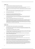
slide 7 – overview of enrichment strategies for phorphopeptides
first trypsinization after enrichment of the peptide
enriched by
— immunoprecipitation
— affinity chromatography
o IMAC
o TiO2
o SIMAC
— chemical derivatization -elimination
Phosphopeptide determination by MS/MS
CID
— Ester phosphate bond is labile and is earlier broken than the peptide bond: formation
of H3PO4 (98 Da) by spontaneous b-elimination.
— Can be dehtected by neutral loss makes use of the 2 MS analyzers that are
connected to each other and scan in a synchronic way
o 1st scans for a peptide with a certain molecular weight
o 2nd scans for a peptide which has lost 98 Da as compared to the molecular
weight of the first scanner
— Phosphotyrosine immonium ions (m/z 216 = a-1 coming from b-1) are relatively easy
formed and are therefore characteristic for the presence of pTyr in the peptide
sequence.
o phosphotyrosine can form immonium ions they appear at the left of the
MSMS spectrum these are the first b-ions that are converted into a-ions
because they loose CO2
o If the peptide has only one tyrosine and you see immonium ion appearing
then you can tell that the peptide was phosphorylated
ECD and ETD we make use of radicals produced by electrons
— Fragmentation occurs exclusively at the peptide backbone and phosphate groups stay
connected to AAs allowing the easy detection of phosphorylation sites (+ 80 Da).
o phosphor-group stays connected with the chain of amino acids
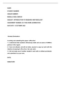
Treatment of the peptides with phosphatase results in a peak that is 80 Da reduced. this
means that the phosphatase did remove the phosphorylating = shift to the left with 80 Da
Example: fraction 4 of b-caseine contains phosphorylated serine that disappears after
phosphatase treatment.
, An example of differential phophoproteomics
people often look at increase/decrease of protein expression levels you can also look at the
increase/decrease of phosphorylation after Dengue virus infection combination of the two
K562 cells macrophage early cell line cells were infected with dengue type 2 virus
cells were lysed
tryptic digestion
peptides were labeled, but in a different way
— first sample: formaldehyde = light form
— second sample: formaldehyde containing 13C = heavy form to differentiate
between the 2 fractions
— Differentially labeled the samples they pooled samples phospho-enrichment in
one group was performed HILIC = chromatography based on hydrophilicity
after that they perform LC-ESI-MS/MS to look at phosphorylation
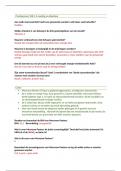
— Graph (slide 10): Total number of proteins exceed 2000
o Fraction that is significantly upregulated and another fraction that is
significantly downregulated
o Phosphoproteins
Fraction of these is upregulated and downregulated
Look at peptides after tryptic digest look at up- and
downregulation of certain phosphosite within the phosphor
proteins
Many phosphoproteins contain different phosphosite so
more phosphorsites than phosphoproteins
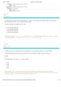
— Slide 11: STRING database: dense network of regulated proteins from “cellular
macromolecule biosynthesis” and “RNA splicing”
o functional enrichment analysis looks for pathways in which your protein
set is enriched in comparison with an ad random set
o Many proteins are connected you get clusters of strongly connected
proteins
the database shows also that many of the proteins are connected
with each other (grey lines) cluster of proteins can be regulated by
the Dengue virus
— Always good to perform a western blot
o Proteins with an astrix are controlled in western blot
— Slide 12: Looking at the phosphor sites and phosphoproteins STRING: dense
network of regulated phosphoproteins from “regulation of transcription” and mRNA
processing”
o Again two clusters
Dengue virus is really trying to influence the biosynthetic process of
macromolecules
You can even predict with kinases where active in the cell. A kinase has always a certain
consensus site given in a set of proteins. A certain kinase phosphorylate always a certain set
of proteins
— on the basis of these sequences, they found out that some sequences were
phosphorylated certain kinases have a preference for certain amino acids/amino
acid sequences this way we can see which kinases are active (by seeing which
amino acid sequences are phosphorylated)
analysis
Posttranslational modifications
Proteins are very regularly phosphorylated, glycosylated, acetylated, ubiquitaned… we can
investigate this on a individual level (e.g. western blot), but it is more difficult to do thison a
proteome-large scale one of the main reasons: not all proteins of a certain type are modificated.
Only a fraction is phosphorylated (e.g. only a small fraction of the STAT protein in the cell is
fosforylated, the other fraction of STAT not)
more sensitive techniques are necessary to see the modified proteins
Cystine (=disulfide) bridge formation
Difference between (–SH)2 and –S-S-: 2 Da very high resolution is necessary.
— if you have a disulfide bridge and if you reduce it 2 protons (1 to a cysteine, 1 to
another cysteine) difference of 2 Da between the oxidated and reduced form
o 2 Da is a low number this is the reason why we need a high resolution
If not: free sulfhydrylgroups can be derivatised, e.g. with para-hydroxy mercury benzoaat
(pHMB) mass shift of 321 Da
— reduced cysteines contain sulfhydrylgroups or thiol groups
— pHMB will make a covalent bound with reduced cysteine
Derivatisation occurs before and after reduction of the protein (with for example B
mercaptoethanol) look for difference in derivatization
trypsinization look at those peptides that are increased with 321 Da sequence the
proteins sequencing shows which cysteines are involved in cystine bridge formation.
Phosphoproteomics
General
Estimation: more than 50% of all proteins is phosphorylated once in their lifetime; more than
100.000 phosphorylation sites.
Big challenge:
— Relative low abundance of phosphorylated proteins only a fraction of the protein
is phosphorylated
— low phosphorylation stoichiometry (many combinations on one protein)
— dynamic regulation (phosphorylation/dephosphorylation)
o proteins can be phosphorylated at that time point that you look at your cell
at another time point, the proteins can be dephosphorylated
this is not fixed
— Negative charge suppresses ionisation low intensities
o with proteomics, we usually work with positively charged proteins
Enrichtment of phosphopeptides
MS is not able to differentiate between phosphorylation and no phosphorylation
Immuno affinity chromatography use antibodies
— Antibodies against pY, pS and pT (last two are less specific; but tyrosine is more
feasible) or against AA sequences that are kinase consensus sites.
o all the kinases in the cell, have their preferences for a certain AA-sequence
if you want to pick up a fosforylated serine that is made by some kind of
kinase, you will make a specific antibody against the phosphorylated
consensus site of that kinase
, o Tyrosine is a bulky protein
o Serine and threonine are less specific
o These AA lie in a consensus site that is recognized by the kinase
— Usually in combination with other methods.
o Antibodies are usually quite difficult molecules to work with not as
specific as you wish.
Immobilised metal affinity chromatography (IMAC)
— Metal ions are chelated with nitrilotriacetic acid (NTA) to beads formation of a
stationairy phase to which negative charged phosphopeptides can bind.
o in the case for iron this is chelated with nitrilotriacetic acid
o they all have an oxygen that is negatively charged will form a salt bridge
with the positively charged iron
o iron = 3+ iron can undergo other electrostatic interactions with phosphor
groups that are reciting on the peptide
— Not very specific (co-purification of non-phosphorylated proteins/peptides with
many acidic AAs)
— Preference for multiple phosphorylated peptides the binding to the beads will be
stronger
— Elution under basic conditions
— Variation: SIMAC (Sequential IMAC chromatography)
3 fractions:
o Flow-through: non-phosphorylated and non-bound phosphorylated peptides
Load column + a neutral buffer everything that doesn’t bind to the
IMAC column will go through. These are not phosphorylated proteins
and non-bind phosphorylated peptides so you will always loose
some phosphorylated peptides
o Acidic elution (less specific): contains non- phosphorylated and
(mono)phosphorylated peptides further analysis on TiO2 column
o Basic elution: contains mostly phosphorylated peptides also the multiple
phosphorylated peptides
Titanium dioxide chromatography
— TiO2 shows affinity for phosphorylated proteins/peptides
— Preference for mono-phosphorylated peptides
— bead covered with titanium dioxide
— Resin in the pipette tip go up and down with solution to let the titanium dioxide
bind to the beads
— the phosphorylated peptides can stick to the titanium also by making an electrostatic
interaction
— changes in pH competition between hydroxyl groups and phosphor groups
SCX = strong cathion exchange chromatography
— At pH 2.7 phosphate containg peptides have one + charge (originating from C-
terminal K or R, all other AAs are protonated). The negative charge of the phosphate
groups (pK=± 1, hence not yet protonated) results in a weak binding to the SCX
column.
o Negative charge of the phosphate group weakens the positivity of the
peptide peptide without phosphate group will bind stronger the column
as compared to the same peptide that contains the phosphorylate
— Because of this weak binding the phosphorylated peptides elute from the column
before the other peptides (e.g. after salt gradient).
— very aspecific, usually in combination with IMAC or TiO 2.
, chemical derivatization (mostly when there is only a limited amount of sample)
remove the phosphor and replace it by a biotin-tag
— Alkalic conditions: b-elimination of phosphate groups on serine and threonine: resp.
dehydroalanine and b-methyldehydroalanine.
— Addition of 1, 2-ethanedithiol results in a di-thiol (Michael addition) to which a biotin
group can be attached after oxidation.
— Biotin group allows for specific purification
o there are beads (these have avidin or streptavidin) that are able to capture
the biotin in of the sample to fish out proteins that were phosphorylated
— for glycosylation there is a similar strategy
slide 7 – overview of enrichment strategies for phorphopeptides
first trypsinization after enrichment of the peptide
enriched by
— immunoprecipitation
— affinity chromatography
o IMAC
o TiO2
o SIMAC
— chemical derivatization -elimination
Phosphopeptide determination by MS/MS
CID
— Ester phosphate bond is labile and is earlier broken than the peptide bond: formation
of H3PO4 (98 Da) by spontaneous b-elimination.
— Can be dehtected by neutral loss makes use of the 2 MS analyzers that are
connected to each other and scan in a synchronic way
o 1st scans for a peptide with a certain molecular weight
o 2nd scans for a peptide which has lost 98 Da as compared to the molecular
weight of the first scanner
— Phosphotyrosine immonium ions (m/z 216 = a-1 coming from b-1) are relatively easy
formed and are therefore characteristic for the presence of pTyr in the peptide
sequence.
o phosphotyrosine can form immonium ions they appear at the left of the
MSMS spectrum these are the first b-ions that are converted into a-ions
because they loose CO2
o If the peptide has only one tyrosine and you see immonium ion appearing
then you can tell that the peptide was phosphorylated
ECD and ETD we make use of radicals produced by electrons
— Fragmentation occurs exclusively at the peptide backbone and phosphate groups stay
connected to AAs allowing the easy detection of phosphorylation sites (+ 80 Da).
o phosphor-group stays connected with the chain of amino acids
Treatment of the peptides with phosphatase results in a peak that is 80 Da reduced. this
means that the phosphatase did remove the phosphorylating = shift to the left with 80 Da
Example: fraction 4 of b-caseine contains phosphorylated serine that disappears after
phosphatase treatment.
, An example of differential phophoproteomics
people often look at increase/decrease of protein expression levels you can also look at the
increase/decrease of phosphorylation after Dengue virus infection combination of the two
K562 cells macrophage early cell line cells were infected with dengue type 2 virus
cells were lysed
tryptic digestion
peptides were labeled, but in a different way
— first sample: formaldehyde = light form
— second sample: formaldehyde containing 13C = heavy form to differentiate
between the 2 fractions
— Differentially labeled the samples they pooled samples phospho-enrichment in
one group was performed HILIC = chromatography based on hydrophilicity
after that they perform LC-ESI-MS/MS to look at phosphorylation
— Graph (slide 10): Total number of proteins exceed 2000
o Fraction that is significantly upregulated and another fraction that is
significantly downregulated
o Phosphoproteins
Fraction of these is upregulated and downregulated
Look at peptides after tryptic digest look at up- and
downregulation of certain phosphosite within the phosphor
proteins
Many phosphoproteins contain different phosphosite so
more phosphorsites than phosphoproteins
— Slide 11: STRING database: dense network of regulated proteins from “cellular
macromolecule biosynthesis” and “RNA splicing”
o functional enrichment analysis looks for pathways in which your protein
set is enriched in comparison with an ad random set
o Many proteins are connected you get clusters of strongly connected
proteins
the database shows also that many of the proteins are connected
with each other (grey lines) cluster of proteins can be regulated by
the Dengue virus
— Always good to perform a western blot
o Proteins with an astrix are controlled in western blot
— Slide 12: Looking at the phosphor sites and phosphoproteins STRING: dense
network of regulated phosphoproteins from “regulation of transcription” and mRNA
processing”
o Again two clusters
Dengue virus is really trying to influence the biosynthetic process of
macromolecules
You can even predict with kinases where active in the cell. A kinase has always a certain
consensus site given in a set of proteins. A certain kinase phosphorylate always a certain set
of proteins
— on the basis of these sequences, they found out that some sequences were
phosphorylated certain kinases have a preference for certain amino acids/amino
acid sequences this way we can see which kinases are active (by seeing which
amino acid sequences are phosphorylated)