MUSCULOSKELETAL CONDITIONS
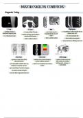
Diagnostic Testing:
X-ray: CT Scan: MRI: Myelogram:
- evaluates structure & - X-ray providing 3D image - use of radio waves & - x-ray study w/ contrast injected into sac
functional changes & joints magnetic field to view soft around nerve root
- identifies soft tissue abnormalities &
- 1 or 2 dimensional views musculoskeletal traumas tissue - CT scan may follow test
- With or without contrast - with or without contrast - detects subtle lesions/injuries
- shows how bone is affecting nerve root
Bone Scan: Dexa Scan: Arthroscopy:
- injection of radio isotope that is - measures bone mineral density w/ - injection of arthroscope into
absorbed by bone minimal radiation exposure joint cavity to dx problems
- Dx: osteomyelitis, metastatic bone Ca, - used for dx of metabolic bone disease with ligaments, cartilage, or
fractures, & avascular necrosis (osteo) joint capsule
- give radioisotope 2 hrs before surgery
- pt needs to be able to lay flat
- increases fluids to get radioisotope out
,Soft Tissue Injuries:
SPRAINS STRAINS
• Injury to ligamentous structures surrounding a joint • An excessive stretching of a muscle or tendon
• Caused by wrenching or twisting motion • Most occur in large muscle groups à lower back,
• Most occur in ankle &/or knee joints calf, hamstring
• Classified according to amt. of ligament fibers tornà • Classified as
- 1st degree: mild - 1st degree: mild or slightly pulled
- 2nd degree: moderate - 2nd degree: moderate or moderately torn
- 3rd degree: se vere (complete tear) - 3rd degree: severely ruptured or torn muscle
• Diagnostic testing and nursing management:
o X-raysà rule out fracture and identify if tissue swelling at site
o MRIà diagnostic for ligament tears
o Ultrasoundà shows diseased or injured ligaments and tendons
o Educateà stretching, balance, strength/conditioning
o s/s: swelling and pain
DISLOCATION SUBLAXATION
• A severe injury to the ligamentous structures that • PARTIAL OR INCOMPLETE displacement of the joint
surround a joint surface
• Results in COMPLETE DISPLACEMENT or separation
of joint surfaces
• Clinical manifestations:
o Pain, tenderness, numbness, tingling
o Loss or reduced function of the injured part
o Swelling
*deformity is the most obvious clinical finding for both*
• Major complications:
o Avascular necrosis à ruptured blood vessel à bone death
o Neurovascular compromise to adjacent tissue à lose pulse to extremity à assess both extremities to compare
o Permanent injury
, o Intra-articular fractures
• Management of dislocation:
o Realignà restore dislocated portion of the joint to its correct anatomical location
§ PRIMARY GOAL
§ The longer the joint is out of place= harder it is to fix
• Muscle tense around joint
o Reduceà closed or open reduction (under local or general anesthesia, or IV conscious sedation)
§ Extremity is immobilized by bracing, splinting, taping, or use of a sling
o Restoreà restore function and use of extremity for optimal function
Carpel Tunnel Syndrome:
• Compression of the median nerveà enters the hand through the carpal tunnel
o Carpal tunnel is formed by ligaments and bones
o Most common upper extremity compression neuropathy
o Associated with repetitive hobbies/occupations
• Clinical manifestations:
o Impaired sensation
o Pain
o Numbness
o Weakness
o Relieved pain with shaking of hands
o Positive Tinel sign and Phalen sign
o Women can have flare ups
§ Menopause, pregnancy, and menstruation
• Treatment modalities:
o Relieve underlying cause
o Educate on identification of risk factors and changed behaviors
o Stop aggravating movements
o Steroid injections
o Surgeryà carpal tunnel release
o Adaptive devices à wrist splints are commonly WORN AT NIGHT to alleviate pressure on median nerve
Fractures:
• Disruption in the continuity of the bone structure
• Majority result from trauma or secondary to disease processes à cancer, osteoporosis
• Classified by complexity and location:
o Can be described using more than one term
o Can have features of multiple classifications
• Classifications include:
o Complete
o Incomplete
o Comminuted
o Closed
o Open