MBZ – juni 2025 69
THE URINARY SYSTEM
Excretion are the processes that remove wastes and excess materials from the body, and there are a few
systems and excretory organs involved:
- The digestive system excretes food residues and wastes produced by the liver
- The respiratory system (lungs) excretes carbon dioxide
- The integumentary system (skin) excretes water and salt
- The urinary system (kidneys) excretes nitrogenous wastes, excess solutes, and water
ORGANS OF THE URINARY SYSTEM
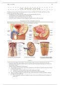
The urinary system consists of kidneys, ureters, urinary bladder and urethra
KIDNEYS
= primary functional organs that produce urine and removes wastes and excess materials from the body
- Located on either side of the vertebral column, near the posterior body wall, the kidneys are the size of a
fist and bean-shaped
- Kidneys maintain homeostasis in many ways:
• They regulate how much water and salt are excreted out of the body to maintain homeostasis of fluid
volume and composition
§ Homeostasis is maintained when water intake equals water output
§ The kidneys either excrete excess water or conserve as much water as possible
• They also regulate nitrogenous wastes and other solutes (like sodium, chloride, potassium, calcium,
hydrogen ions and creatinine)
§ Protein metabolism produces nitrogenous wastes, like ammonia (NH₃) during the breakdown of
amino acids
,MBZ – juni 2025 70
§ Ammonia is toxic to the body, so the liver detoxifies it by converting it into urea
§ The urea is then transported from the liver to the kidneys, where it is excreted through urine
• They secrete an enzyme involved in the control of blood volume and blood pressure
• They maintain acid-base balance and blood pH
• They control production of RBC
• They activate an inactive form of vitamin D
INTERNAL STRUCTURE
- The renal artery connects to the aorta, while the renal vein connects to the inferior vena cava
- The medulla or renal pyramids are pyramid-shaped zones of dense tissue
- The area around the medulla is called the cortex
- At the center of the kidney is a hollow space, the renal pelvis, where urine collects after it is formed
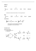
- Nephrons are tubular structures that produce urine
• Each kidney has about 1 million of them
• An individual nephron consists of a tubule (a thin, hollow tube of epithelial cells) and associated blood
vessels
§ The glomerular capsule encloses a network of capillaries called the glomerulus
§ Proximal tubule: nearest to the capsule, extends from the cortex to the medulla
§ Loop of Henle: U-shaped structure with a descending and ascending limb
§ Distal tubule: further from the capsule
• Thousands of nephrons join into a collecting duct, through which urine is delivered to the renal pelvis
• They remove 180 liters of fluid from the blood daily and return most of it, leaving behind only a small
amount as urine
Distal tubule
Glomerular capsule
Proximal tubule
Descending limb
Loop of Henle
Ascending limb
BLOOD SUPPLY TO NEPHRONS
The renal artery and renal vein branch many times to deliver blood to each glomerulus
- The afferent arteriole enters a glomerular capsule and then divides into a network of capillaries that forms
the glomerulus
• Glomerular capillaries filter plasma fluid and solutes from the blood into the capsular space
- The efferent arteriole carries filtered blood away from the glomerulus and divides again into another
capillary network that surrounds the proximal and distal tubules, called peritubular capillaries
,MBZ – juni 2025 71
• Peritubular capillaries remove water, ions and nutrients, which are reabsorbed by the proximal and
distal tubules
• Efferent arterioles of a few nephrons descend into the medulla and divide into long, thin capillaries
called the vasa recta, which supply the loop of Henle and collecting duct
FORMATION OF URINE
The urinary system maintains fluid balance and homeostasis by selectively filtering, reabsorbing, and secreting
substances in order to retain essential nutrients, while excess ions and wastes are excreted
GLOMERULAR FILTRATION
Plasma fluid and solutes (but not proteins or blood cells) pass from the glomerulus into the glomerular capsule
- The filtration barrier consists of capillary cells and podocyte (modified epithelial cells that surround the
capillaries) and functions like a sieve, allowing water and small solutes to pass through while preventing
large proteins and blood cells from entering the filtrate
- The kidneys filter approximately 180 liters of plasma fluid per day, but only about 1,5 liters of urine are
excreted, highlighting their efficiency in reabsorbing useful substances
- The process is driven by high blood pressure in the glomerular capillaries, which is maintained because the
efferent arteriole is narrower than the afferent arteriole, creating resistance and pressure buildup
- Glomerular filtration rate is regulated in two main ways:
• Under normal conditions, local feedback mechanisms adjust the afferent arteriole diameter to keep
filtration rates stable
• During stress (like blood loss or intense exercise), the sympathetic nervous system constricts the
arterioles, reducing filtration and urine production to conserve fluids and redirect blood to essential
organs
- Disruptions in the filtration barrier can lead to proteinuria, where proteins leak into the urine
• Persistent proteinuria may indicate kidney damage due to high blood pressure or toxins
• Temporary proteinuria can occur after strenuous exercise
• Exercise-induced proteinuria is harmless and usually resolves within a day
, MBZ – juni 2025 72
TUBULAR REABSORPTION
Most fluids and solutes are reabsorbed from the tubule into the peritubular capillaries or vasa recta
- As the filtrate moves through the tubule
• Essential nutrients like glucose, amino acids, and bicarbonate, and more than 99% of water and sodium
are reabsorbed
• About 50% of urea is also reabsorbed
• Waste products like creatinine are not reabsorbed at all
• The final urine contains only the necessary amount of water, sodium, urea, and trace amounts of other
waste products to maintain body balance
- Most reabsorption occurs automatically in the proximal tubule (65–70% of water) and the loop of Henle
(25%), but the final adjustment of how much water the body actually retains or excretes happens in the
distal tubule and collecting duct (even though they absorb less than 10%)
- For substances to be reabsorbed, they must pass through the
epithelial cells lining the proximal tubule, which have microvilli
that increase surface area for absorption
- The entire process depends on one key active step:
1. Active transport of sodium (Na⁺) out of the proximal tubule
cell, using ATP for energy
2. Sodium then diffuses into the capillary, lowering sodium
levels inside the tubule cell
3. This creates a concentration gradient, allowing more sodium
to passively move from the tubule (filtrate) into the tubule
cell
4. Chloride (Cl⁻) follows sodium to maintain electrical balance
5. Water follows sodium and chloride due to osmosis, leading to water
reabsorption
6. The sodium gradient powers the co-transport of glucose and amino
acids into the tubule cell
7. Glucose and amino acids then diffuse into the blood, completing
reabsorption
ð The driving force behind reabsorption is the active transport of sodium
• It ensures the efficient absorption of water, chloride, glucose, and
amino acids
• In the distal tubule, reabsorption continues under hormonal control,
helping fine-tune the body’s water and electrolyte balance
THE URINARY SYSTEM
Excretion are the processes that remove wastes and excess materials from the body, and there are a few
systems and excretory organs involved:
- The digestive system excretes food residues and wastes produced by the liver
- The respiratory system (lungs) excretes carbon dioxide
- The integumentary system (skin) excretes water and salt
- The urinary system (kidneys) excretes nitrogenous wastes, excess solutes, and water
ORGANS OF THE URINARY SYSTEM
The urinary system consists of kidneys, ureters, urinary bladder and urethra
KIDNEYS
= primary functional organs that produce urine and removes wastes and excess materials from the body
- Located on either side of the vertebral column, near the posterior body wall, the kidneys are the size of a
fist and bean-shaped
- Kidneys maintain homeostasis in many ways:
• They regulate how much water and salt are excreted out of the body to maintain homeostasis of fluid
volume and composition
§ Homeostasis is maintained when water intake equals water output
§ The kidneys either excrete excess water or conserve as much water as possible
• They also regulate nitrogenous wastes and other solutes (like sodium, chloride, potassium, calcium,
hydrogen ions and creatinine)
§ Protein metabolism produces nitrogenous wastes, like ammonia (NH₃) during the breakdown of
amino acids
,MBZ – juni 2025 70
§ Ammonia is toxic to the body, so the liver detoxifies it by converting it into urea
§ The urea is then transported from the liver to the kidneys, where it is excreted through urine
• They secrete an enzyme involved in the control of blood volume and blood pressure
• They maintain acid-base balance and blood pH
• They control production of RBC
• They activate an inactive form of vitamin D
INTERNAL STRUCTURE
- The renal artery connects to the aorta, while the renal vein connects to the inferior vena cava
- The medulla or renal pyramids are pyramid-shaped zones of dense tissue
- The area around the medulla is called the cortex
- At the center of the kidney is a hollow space, the renal pelvis, where urine collects after it is formed
- Nephrons are tubular structures that produce urine
• Each kidney has about 1 million of them
• An individual nephron consists of a tubule (a thin, hollow tube of epithelial cells) and associated blood
vessels
§ The glomerular capsule encloses a network of capillaries called the glomerulus
§ Proximal tubule: nearest to the capsule, extends from the cortex to the medulla
§ Loop of Henle: U-shaped structure with a descending and ascending limb
§ Distal tubule: further from the capsule
• Thousands of nephrons join into a collecting duct, through which urine is delivered to the renal pelvis
• They remove 180 liters of fluid from the blood daily and return most of it, leaving behind only a small
amount as urine
Distal tubule
Glomerular capsule
Proximal tubule
Descending limb
Loop of Henle
Ascending limb
BLOOD SUPPLY TO NEPHRONS
The renal artery and renal vein branch many times to deliver blood to each glomerulus
- The afferent arteriole enters a glomerular capsule and then divides into a network of capillaries that forms
the glomerulus
• Glomerular capillaries filter plasma fluid and solutes from the blood into the capsular space
- The efferent arteriole carries filtered blood away from the glomerulus and divides again into another
capillary network that surrounds the proximal and distal tubules, called peritubular capillaries
,MBZ – juni 2025 71
• Peritubular capillaries remove water, ions and nutrients, which are reabsorbed by the proximal and
distal tubules
• Efferent arterioles of a few nephrons descend into the medulla and divide into long, thin capillaries
called the vasa recta, which supply the loop of Henle and collecting duct
FORMATION OF URINE
The urinary system maintains fluid balance and homeostasis by selectively filtering, reabsorbing, and secreting
substances in order to retain essential nutrients, while excess ions and wastes are excreted
GLOMERULAR FILTRATION
Plasma fluid and solutes (but not proteins or blood cells) pass from the glomerulus into the glomerular capsule
- The filtration barrier consists of capillary cells and podocyte (modified epithelial cells that surround the
capillaries) and functions like a sieve, allowing water and small solutes to pass through while preventing
large proteins and blood cells from entering the filtrate
- The kidneys filter approximately 180 liters of plasma fluid per day, but only about 1,5 liters of urine are
excreted, highlighting their efficiency in reabsorbing useful substances
- The process is driven by high blood pressure in the glomerular capillaries, which is maintained because the
efferent arteriole is narrower than the afferent arteriole, creating resistance and pressure buildup
- Glomerular filtration rate is regulated in two main ways:
• Under normal conditions, local feedback mechanisms adjust the afferent arteriole diameter to keep
filtration rates stable
• During stress (like blood loss or intense exercise), the sympathetic nervous system constricts the
arterioles, reducing filtration and urine production to conserve fluids and redirect blood to essential
organs
- Disruptions in the filtration barrier can lead to proteinuria, where proteins leak into the urine
• Persistent proteinuria may indicate kidney damage due to high blood pressure or toxins
• Temporary proteinuria can occur after strenuous exercise
• Exercise-induced proteinuria is harmless and usually resolves within a day
, MBZ – juni 2025 72
TUBULAR REABSORPTION
Most fluids and solutes are reabsorbed from the tubule into the peritubular capillaries or vasa recta
- As the filtrate moves through the tubule
• Essential nutrients like glucose, amino acids, and bicarbonate, and more than 99% of water and sodium
are reabsorbed
• About 50% of urea is also reabsorbed
• Waste products like creatinine are not reabsorbed at all
• The final urine contains only the necessary amount of water, sodium, urea, and trace amounts of other
waste products to maintain body balance
- Most reabsorption occurs automatically in the proximal tubule (65–70% of water) and the loop of Henle
(25%), but the final adjustment of how much water the body actually retains or excretes happens in the
distal tubule and collecting duct (even though they absorb less than 10%)
- For substances to be reabsorbed, they must pass through the
epithelial cells lining the proximal tubule, which have microvilli
that increase surface area for absorption
- The entire process depends on one key active step:
1. Active transport of sodium (Na⁺) out of the proximal tubule
cell, using ATP for energy
2. Sodium then diffuses into the capillary, lowering sodium
levels inside the tubule cell
3. This creates a concentration gradient, allowing more sodium
to passively move from the tubule (filtrate) into the tubule
cell
4. Chloride (Cl⁻) follows sodium to maintain electrical balance
5. Water follows sodium and chloride due to osmosis, leading to water
reabsorption
6. The sodium gradient powers the co-transport of glucose and amino
acids into the tubule cell
7. Glucose and amino acids then diffuse into the blood, completing
reabsorption
ð The driving force behind reabsorption is the active transport of sodium
• It ensures the efficient absorption of water, chloride, glucose, and
amino acids
• In the distal tubule, reabsorption continues under hormonal control,
helping fine-tune the body’s water and electrolyte balance