JACKLINE
EKG Kaiser Study Guide With Questions And 100% ALL SURE ANSWERS
Terms in this set (48)
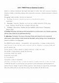
Cardiac Conduction Pathway System SA node> AV node> Bundle of His> Left and Right bundle branches> Purkinjie fibers
♥ Natural: SA Node = 60-100 beats/minute
Pacemakers of the heart ♥ Backup: AV Node = 40-60 beats/minute
♥ Backup: Purkinje Fibers (ventricles) = 20-40 beats/minute
• Intrinsic Pacemaker of the heart
• Rate 60-100 beats/min
SA Node • RA (Right Atrium), close to SVC (Superior Vena Cava)
• Blood supply from RCA (Right Coronary Artery) & LCA
(Left Coronary Artery)
EKG Kaiser Study Guide
1/26
, Transmits impulses from the SA node to the AV node
through the RA & LA
Interatrial/Internodal Tracts
EKG Kaiser Study Guide
2/26
, 10/23/24, 1:36 PM
• Slows conduction (40-60 beats/min)
• Physiologic delay allows atrial kick on floor of RA near
AV node
tricuspid valve
Bundle of cardiac muscle fibers that conducts the
electrical impulses from the AV node in the right atrium
Bundle of His
to the septum between the ventricles and then to the
left and right ventricles.
Right and Left (Left anterior & Left posterior fascicles)
bundle branches
Fibers from Bundle Branches imbedded into the
ventricle walls.
Purkinjie Fibers
Depolarization – electrical excitation of the cell membrane, normally followed by mechanical
contraction
Depolarization vs Repolarization
Repolarization – return of cell membrane to its resting state, normally followed by mechanical
relaxation
♥ The heart has two activities that are performed rhythmically: electrical activity followed by
mechanical activity
Electrical and mechanical activity of the heart ♥ Electrical activity always precedes mechanical activity
♥ It is possible to have electrical activity without mechanical response
♥ Always check the patient - Do Not Depend on the Machine!!!
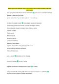
♥ A standard ECG is printed at 25mm per second or 25
small squares per second. Since one second divided by
25 small boxes, then each 1 mm box = 0.04 seconds.
The larger boxes indicated by the heavier lines are
equal to 0.20 seconds.
Measurement of the boxes in the ekg
♥ Voltage is measured along the vertical axis and is
expressed in millivolts (mV). The standard calibration is
that a 1 mV signal produces a 10-mm deflection (0.1
mV=1mm). Simply put 10 small squares vertically is
equal to 1 millivolt
EKG Kaiser Study Guide
3/26
EKG Kaiser Study Guide With Questions And 100% ALL SURE ANSWERS
Terms in this set (48)
Cardiac Conduction Pathway System SA node> AV node> Bundle of His> Left and Right bundle branches> Purkinjie fibers
♥ Natural: SA Node = 60-100 beats/minute
Pacemakers of the heart ♥ Backup: AV Node = 40-60 beats/minute
♥ Backup: Purkinje Fibers (ventricles) = 20-40 beats/minute
• Intrinsic Pacemaker of the heart
• Rate 60-100 beats/min
SA Node • RA (Right Atrium), close to SVC (Superior Vena Cava)
• Blood supply from RCA (Right Coronary Artery) & LCA
(Left Coronary Artery)
EKG Kaiser Study Guide
1/26
, Transmits impulses from the SA node to the AV node
through the RA & LA
Interatrial/Internodal Tracts
EKG Kaiser Study Guide
2/26
, 10/23/24, 1:36 PM
• Slows conduction (40-60 beats/min)
• Physiologic delay allows atrial kick on floor of RA near
AV node
tricuspid valve
Bundle of cardiac muscle fibers that conducts the
electrical impulses from the AV node in the right atrium
Bundle of His
to the septum between the ventricles and then to the
left and right ventricles.
Right and Left (Left anterior & Left posterior fascicles)
bundle branches
Fibers from Bundle Branches imbedded into the
ventricle walls.
Purkinjie Fibers
Depolarization – electrical excitation of the cell membrane, normally followed by mechanical
contraction
Depolarization vs Repolarization
Repolarization – return of cell membrane to its resting state, normally followed by mechanical
relaxation
♥ The heart has two activities that are performed rhythmically: electrical activity followed by
mechanical activity
Electrical and mechanical activity of the heart ♥ Electrical activity always precedes mechanical activity
♥ It is possible to have electrical activity without mechanical response
♥ Always check the patient - Do Not Depend on the Machine!!!
♥ A standard ECG is printed at 25mm per second or 25
small squares per second. Since one second divided by
25 small boxes, then each 1 mm box = 0.04 seconds.
The larger boxes indicated by the heavier lines are
equal to 0.20 seconds.
Measurement of the boxes in the ekg
♥ Voltage is measured along the vertical axis and is
expressed in millivolts (mV). The standard calibration is
that a 1 mV signal produces a 10-mm deflection (0.1
mV=1mm). Simply put 10 small squares vertically is
equal to 1 millivolt
EKG Kaiser Study Guide
3/26