Cell Structure Notes
2.1.1 Microscopes
- Magnification = number of times larger an image appears compared with the size of the object
Microscopes produce linear magnification: magnification of x100 is 100x wider and 100x longer
than it actually is
- Resolution = the clarity of an image, how an optical instrument can show fine detail clearly
Smallest distance between two points that can still be seen as two points
- Total magnification = magnification of objective lens x magnifying power of the eyepiece lens
- Electron micrograph = photograph of an image seen using an electron microscope
- Magnification = number of times larger an image appears compared with the size of the object
- Organelles = small structures within cells, each with a specific function
- Photomicrograph = photograph of an image seen using an optical microscope
- Resolution = the clarity of an image, higher resolution = clearer image
- Eyes, optical microscopes and electron microscopes are all optical instruments
- The logarithmic scale goes up in steps – each 10-fold increase of the previous and is used to
show which organisms/organelles can be seen by which optical instrument
Op#cal/light microscopes
- Optical (light) microscopes are used because they are:
a) relatively cheap
b) easy to use
c) portable to use in the field and labs
d) able to study whole living specimens
- Allow magnification up to x1500 or x2000 to see some larger sub-cellular structures in cells but
due to limited resolution cant magnify any higher and still be clear
- Objective lens 4x/10x/40x/100x
- Eyepiece lens 10x/15x
- Produces 2D images
- Visible light wavelength 400-700nm, structures closer together than 200nm (400/2) appear as 1
object, e.g. Ribosomes have a 20nm diameter so can’t be seen
, Laser scanning microscopes/confocal microscopes
- Use a laser light to scan an object point by point
- A computer assembles the pixel information onto one 3D image – displayed on the screen
- High resolution
- High contrast
- 1000x magnification
- Depth selectivity of the microscopes can focus on internal structures at different depths – can
therefore study clearly whole living organisms and cells
- Used in medicine to give a fast diagnosis and more effective treatment as a result
- Used in many areas of biological research
- Expensive
Electron microscopes
- Beam of fast-travelling electrons (wavelength 0.004nm) fired from a cathode and focused by
magnets (not glass lenses) onto a screen or photographic plate
- Much greater resolution than light microscopes because electrons have a wavelength 125,000x
smaller than visible
- Clear and highly magnified images
- Disadvantages compared to light microscopes:
a) Highly skilled process, needs training
b) Expensive
c) Requires killing the specimen
d) Specimen has to be put in a vacuum
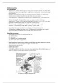
Transmission electron microscopes (TEM)
- Specimen is chemically fixed by being a) dehydrated and b) stained with metal salts
- Beam of electrons passes through a specimen
- Electrons (wavelength 0.004nm) form a 2D black and white (grey scale) image – when
photographed called an electron micrograph
- Magnification of up to 2,000,000x, with ongoing development of up to 50,000,000x
- Shows internal organelles and structures of only dead organisms (due to the dehydrating and
chemical staining)
Scanning electron microscopes (SEM)
- Electrons (0.004nm wavelength) don’t pass through the specimen but instead cause secondary
electrons to bounce off it to be focused onto a screen
- The specimen is whole and only the surface can be examined
- 3D image produced
- Magnification from 15x to 200,000x
- Black and white image, can add false colour from software
- Specimen placed in a vacuum and coated with a film of metal (potentially harmful to the user)
2.1.2 Slides and micrographs
- Light microscopes can be used to view living organisms e.g. Paramecium and Amoeba, smear
preparations of human blood and cheek cells, thin sections of animal/plant/fungal tissue e.g.
bone, muscle, root etc
- Many specimens are colourless and transparent, in order to see them:
a) Use light inference rather than light absorption microscope to produce a clear image
b) Use a dark background so the specimen shows up
c) Adjust the iris diaphragm to reduce the illumination of the specimen
2.1.1 Microscopes
- Magnification = number of times larger an image appears compared with the size of the object
Microscopes produce linear magnification: magnification of x100 is 100x wider and 100x longer
than it actually is
- Resolution = the clarity of an image, how an optical instrument can show fine detail clearly
Smallest distance between two points that can still be seen as two points
- Total magnification = magnification of objective lens x magnifying power of the eyepiece lens
- Electron micrograph = photograph of an image seen using an electron microscope
- Magnification = number of times larger an image appears compared with the size of the object
- Organelles = small structures within cells, each with a specific function
- Photomicrograph = photograph of an image seen using an optical microscope
- Resolution = the clarity of an image, higher resolution = clearer image
- Eyes, optical microscopes and electron microscopes are all optical instruments
- The logarithmic scale goes up in steps – each 10-fold increase of the previous and is used to
show which organisms/organelles can be seen by which optical instrument
Op#cal/light microscopes
- Optical (light) microscopes are used because they are:
a) relatively cheap
b) easy to use
c) portable to use in the field and labs
d) able to study whole living specimens
- Allow magnification up to x1500 or x2000 to see some larger sub-cellular structures in cells but
due to limited resolution cant magnify any higher and still be clear
- Objective lens 4x/10x/40x/100x
- Eyepiece lens 10x/15x
- Produces 2D images
- Visible light wavelength 400-700nm, structures closer together than 200nm (400/2) appear as 1
object, e.g. Ribosomes have a 20nm diameter so can’t be seen
, Laser scanning microscopes/confocal microscopes
- Use a laser light to scan an object point by point
- A computer assembles the pixel information onto one 3D image – displayed on the screen
- High resolution
- High contrast
- 1000x magnification
- Depth selectivity of the microscopes can focus on internal structures at different depths – can
therefore study clearly whole living organisms and cells
- Used in medicine to give a fast diagnosis and more effective treatment as a result
- Used in many areas of biological research
- Expensive
Electron microscopes
- Beam of fast-travelling electrons (wavelength 0.004nm) fired from a cathode and focused by
magnets (not glass lenses) onto a screen or photographic plate
- Much greater resolution than light microscopes because electrons have a wavelength 125,000x
smaller than visible
- Clear and highly magnified images
- Disadvantages compared to light microscopes:
a) Highly skilled process, needs training
b) Expensive
c) Requires killing the specimen
d) Specimen has to be put in a vacuum
Transmission electron microscopes (TEM)
- Specimen is chemically fixed by being a) dehydrated and b) stained with metal salts
- Beam of electrons passes through a specimen
- Electrons (wavelength 0.004nm) form a 2D black and white (grey scale) image – when
photographed called an electron micrograph
- Magnification of up to 2,000,000x, with ongoing development of up to 50,000,000x
- Shows internal organelles and structures of only dead organisms (due to the dehydrating and
chemical staining)
Scanning electron microscopes (SEM)
- Electrons (0.004nm wavelength) don’t pass through the specimen but instead cause secondary
electrons to bounce off it to be focused onto a screen
- The specimen is whole and only the surface can be examined
- 3D image produced
- Magnification from 15x to 200,000x
- Black and white image, can add false colour from software
- Specimen placed in a vacuum and coated with a film of metal (potentially harmful to the user)
2.1.2 Slides and micrographs
- Light microscopes can be used to view living organisms e.g. Paramecium and Amoeba, smear
preparations of human blood and cheek cells, thin sections of animal/plant/fungal tissue e.g.
bone, muscle, root etc
- Many specimens are colourless and transparent, in order to see them:
a) Use light inference rather than light absorption microscope to produce a clear image
b) Use a dark background so the specimen shows up
c) Adjust the iris diaphragm to reduce the illumination of the specimen