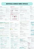
UNIT 21
Medical Physics Application
Radiation use in medical diagnosis and treatment
MEHREEN LATIF 12F
,INTRO
In learning aim A I'm going to explore the principles, production, uses and benefits of
non-ionising instrumentation techniques in medical applications and make a
handbook of it including examples.
1. NON-IONISING RADIATION-
Non-ionising radiation is a type of electromagnetic radiation that is incapable of
ionising atoms or molecules because it lacks the necessary energy. This kind of
radiation is regarded as less dangerous than ionising radiation, which can harm DNA
and raise the risk of developing cancer.
● MRI (Magnetic Resonance Imaging)
● LASERS
● ULTRASOUND
● IRT (Infrared Thermography)
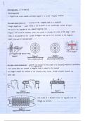
MRI (Magnetic Resonance Imaging)-
It is a medical imaging technique that uses a powerful magnetic field, radio waves,
and a computer to produce detailed images of the inside of the body.
● Production and Principle
Powerful magnets are used to create a strong magnetic field, which aligns the
protons in the body's tissues, in order to produce MRI in non-ionizing radiation. The
protons are then made to emit signals using radio waves, which are subsequently
picked up by a receiver and processed by a computer to provide images of the
body's internal organs.
To produce images, MRI devices
combine radio frequency coils,
gradient magnets, and powerful static
magnets. The protons in the body's
tissues are aligned by the magnets'
homogeneous magnetic field. The
gradient magnets produce a changing
magnetic field that makes it possible
to precisely localise the signal coming
from various bodily parts.
However, due to safety concerns,
some individuals with specific metal
,implants, such as pacemakers or cochlear implants, may not be allowed to undergo
an MRI scan.
● Procedure
The entire MRI procedure typically takes 30-60 minutes, depending on the area of
the body being imaged and whether or not contrast material is used.
Before the MRI process, you will be requested to change into a hospital gown and
take off any jewellery or timepieces that are made of metal. A screening
questionnaire will also be given to you to make sure you don't have any metal
implants or other conditions that would make an MRI dangerous.
You will lie down on a little table that glides into the MRI scanner for positioning. The
scanner will be positioned over the part of your body that needs to be imaged.
When you are positioned correctly, the MRI technician will begin the scan. The
scanner will make loud tapping and knocking noises while it scans your body for
images. To avoid any image blurring during the scan, it's crucial to remain steady.
To help emphasise certain tissues or blood arteries on the MRI images, a contrast
substance may occasionally be injected into a vein.
The technician will then remove any IV lines or other monitoring devices after the
scan is finished and slip you out of the scanner.
The radiologist will review the images following the scan to determine whether any
additional tests are necessary. The radiologist will then draft a report.
● Uses
Soft tissue injury diagnosis: MRI is particularly helpful for identifying injuries to soft
tissues such as tendons, ligaments, and cartilage.
, Imaging of the brain: MRI is routinely used to get precise images of the brain.
Conditions including brain tumours, stroke, multiple sclerosis, and dementia can all
be diagnosed with its assistance. In research investigations and in the diagnosis of
some neurological illnesses, functional MRI (fMRI) is a specialised technology that
can be used to assess brain activity.
MRI is a useful instrument for capturing images of the spinal cord and its
surroundings. It can be used to identify spine malignancies, spinal cord injury, and
degenerative diseases such spinal stenosis.
Heart imaging: MRI scans of the heart and blood arteries are possible. It is especially
helpful for assessing heart health, blood flow, and the presence of obstructions or
anomalies in the blood arteries.
Imaging of the Abdomen: MRI can give fine-grained images of the organs in the
abdomen, including the liver, pancreas, and kidneys. It can be used to identify
diseases such liver illness, cysts, and tumours.
Breast imaging: MRI and mammography are occasionally used to check for breast
cancer in high-risk patients. Moreover, it can be utilised to assess any anomalies or
breast masses.
Imaging of the musculoskeletal system: MRI is helpful for imaging the bones, joints,
and muscles. It can aid in the diagnosis of ailments like arthritis, bone cancers, and
herniated discs.
● Examples
Brain MRI: This popular MRI technique is used to visualise the brain and identify
diseases like tumours, strokes, and multiple sclerosis.
Heart MRI: This kind of MRI is used to visualise the heart and identify diseases such
congenital heart defects, heart failure, and heart disease.
Using an abdominal MRI, doctors may view abdominal organs including the liver,
pancreas, and kidneys and identify diseases like tumours and cysts.
● Benefits and limitations
Benefits:
Ionising radiation is not used in MRI, which is one of its main advantages over CT
and X-ray imaging. This indicates that there is no chance of radiation exposure or
cell harm.
Medical Physics Application
Radiation use in medical diagnosis and treatment
MEHREEN LATIF 12F
,INTRO
In learning aim A I'm going to explore the principles, production, uses and benefits of
non-ionising instrumentation techniques in medical applications and make a
handbook of it including examples.
1. NON-IONISING RADIATION-
Non-ionising radiation is a type of electromagnetic radiation that is incapable of
ionising atoms or molecules because it lacks the necessary energy. This kind of
radiation is regarded as less dangerous than ionising radiation, which can harm DNA
and raise the risk of developing cancer.
● MRI (Magnetic Resonance Imaging)
● LASERS
● ULTRASOUND
● IRT (Infrared Thermography)
MRI (Magnetic Resonance Imaging)-
It is a medical imaging technique that uses a powerful magnetic field, radio waves,
and a computer to produce detailed images of the inside of the body.
● Production and Principle
Powerful magnets are used to create a strong magnetic field, which aligns the
protons in the body's tissues, in order to produce MRI in non-ionizing radiation. The
protons are then made to emit signals using radio waves, which are subsequently
picked up by a receiver and processed by a computer to provide images of the
body's internal organs.
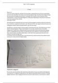
To produce images, MRI devices
combine radio frequency coils,
gradient magnets, and powerful static
magnets. The protons in the body's
tissues are aligned by the magnets'
homogeneous magnetic field. The
gradient magnets produce a changing
magnetic field that makes it possible
to precisely localise the signal coming
from various bodily parts.
However, due to safety concerns,
some individuals with specific metal
,implants, such as pacemakers or cochlear implants, may not be allowed to undergo
an MRI scan.
● Procedure
The entire MRI procedure typically takes 30-60 minutes, depending on the area of
the body being imaged and whether or not contrast material is used.
Before the MRI process, you will be requested to change into a hospital gown and
take off any jewellery or timepieces that are made of metal. A screening
questionnaire will also be given to you to make sure you don't have any metal
implants or other conditions that would make an MRI dangerous.
You will lie down on a little table that glides into the MRI scanner for positioning. The
scanner will be positioned over the part of your body that needs to be imaged.
When you are positioned correctly, the MRI technician will begin the scan. The
scanner will make loud tapping and knocking noises while it scans your body for
images. To avoid any image blurring during the scan, it's crucial to remain steady.
To help emphasise certain tissues or blood arteries on the MRI images, a contrast
substance may occasionally be injected into a vein.
The technician will then remove any IV lines or other monitoring devices after the
scan is finished and slip you out of the scanner.
The radiologist will review the images following the scan to determine whether any
additional tests are necessary. The radiologist will then draft a report.
● Uses
Soft tissue injury diagnosis: MRI is particularly helpful for identifying injuries to soft
tissues such as tendons, ligaments, and cartilage.
, Imaging of the brain: MRI is routinely used to get precise images of the brain.
Conditions including brain tumours, stroke, multiple sclerosis, and dementia can all
be diagnosed with its assistance. In research investigations and in the diagnosis of
some neurological illnesses, functional MRI (fMRI) is a specialised technology that
can be used to assess brain activity.
MRI is a useful instrument for capturing images of the spinal cord and its
surroundings. It can be used to identify spine malignancies, spinal cord injury, and
degenerative diseases such spinal stenosis.
Heart imaging: MRI scans of the heart and blood arteries are possible. It is especially
helpful for assessing heart health, blood flow, and the presence of obstructions or
anomalies in the blood arteries.
Imaging of the Abdomen: MRI can give fine-grained images of the organs in the
abdomen, including the liver, pancreas, and kidneys. It can be used to identify
diseases such liver illness, cysts, and tumours.
Breast imaging: MRI and mammography are occasionally used to check for breast
cancer in high-risk patients. Moreover, it can be utilised to assess any anomalies or
breast masses.
Imaging of the musculoskeletal system: MRI is helpful for imaging the bones, joints,
and muscles. It can aid in the diagnosis of ailments like arthritis, bone cancers, and
herniated discs.
● Examples
Brain MRI: This popular MRI technique is used to visualise the brain and identify
diseases like tumours, strokes, and multiple sclerosis.
Heart MRI: This kind of MRI is used to visualise the heart and identify diseases such
congenital heart defects, heart failure, and heart disease.
Using an abdominal MRI, doctors may view abdominal organs including the liver,
pancreas, and kidneys and identify diseases like tumours and cysts.
● Benefits and limitations
Benefits:
Ionising radiation is not used in MRI, which is one of its main advantages over CT
and X-ray imaging. This indicates that there is no chance of radiation exposure or
cell harm.