Topic 1: epithelial cells and diseases
Day 1: February 7th, Ben Giepmans
10μm = cell, 100nm = mitochondria, 10nm = large proteins/antibodies, 1nm = ATP
GFP = green fluorescent protein = few nanometers to follow cells to understand the dynamics
5-10nm molecule determines what is going on in terms of drug development.
We’re mostly looking at dead cells under the microscope
Desmosomes and blisters diseases → sometimes babies are born with their skin
stripped of because of a genetic defect (because there are hardly any pathogens from
the outside present in the placenta).
We can screen for this before the baby is born (e.g.: Down syndrome, Huntington) to
prevent the more than 50% miscarriage, not compatible with life in the first weeks.
PCR test = polymerase chain reaction, speeds up our understanding in biomedicine
(hence is one of the biggest discoveries in a century in this field).
Diseases can be caused by several things:
• Pathogens (bacteria, viruses, etc.) and other environmental factors
• Genetic defects can also cause diseases
• Own immune system sometimes causes disease: autoimmune disease (could also be genetic)!
• Mostly covered in this course
• Diabetes type 2 could be caused by either one of these three factors in several ways.
o Environmental: obesity due to lifestyle
o Genetic: diabetes can be genetic
o Autoimmune: cells are resistant to own produced insulin
Baseline of this course: if we understand diseases, we can try to prevent / cure them
Hypothesis: disease is cured → people get older → people produce more offspring → more people are
carrier of the diseases.
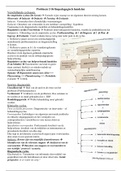
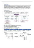
Cell-cell junctions and proteins (discovered by light/electron
microscope (EM))
• Tight junctions are important because they form a barrier
for certain stuff that wants to enter/leave the cell. They
are on top of cells and in between the cells.
• Two barriers:
o One from outside to in: this way, no
bacteria/toxins can enter the cell.
o One to form two compartments on the inside of
the cell.
• Other cell-cell junctions: adherens junctions (functional
name: keeps the cells together, more important for
cellular structure), desmosomes (keep cells together,
more important for tissue structure), gap junctions (are
further apart to form cell to cell channels)
,Tight junctions: made up of transmembrane proteins (e.g., occludin, claudins, junction adhesion
molecules (=JAMs). Several strands of proteins can be observed.
• Functions:
o (1) Barrier function,
o (2) Fence function
o (3) Signal transduction function
• Claudins: big family (20-30 different ones), if there is a problem with a certain claudin, it will
typically affect a specific kind of tissue
o E.g., claudin-14: hereditary deafness
• Tight junctions can be found all across the cell.
Adherens junctions: zona adherens: adhesion belt with cadherins
important for cellular structure.
• Zip cells together for cell strength → cytoskeleton anchoring
• Classical cadherins are named after their location.
o E-cadherin: epithelia
o N-cadherin: neurons, heart, skeletal muscle, and
fibroblasts
o P-cadherin: placenta, epidermis, breast epithelium
o VE-cadherin: endothelial cells.
• Cadherins are transmembrane proteins that are dependent
on calcium, which is needed to be able to form a dimer.
• Catenins are associated to form a bridge to link
transmembrane cadherins to actin cytoskeleton to provide
strength.
,Desmosome: has similar cadherins as adherens junctions but link intermediate filaments instead of actin.
Going from one cell to another they form strong links.
• There are linker proteins to link one desmosome to another providing tissue strength
• Pathology: defects in desmosome gene/protein leads to a certain disease (e.g., autoantibodies)
• Hemidesmosomes: integrins in extracellular matrix
• Cell-cell adhesion between cells: anchor cytoskeleton for strength in tissue (dense structures)!
Gap junctions: large array of channels going from one cell to another (connect cells, what happens in one
cell, automatically happens in the next cell → one cell excited, K channels leads to excitement of the next
cell).
• Allows diffusion of small molecules (<1nm: ATP, calcium, glucose):
ions, nutrients, amino acids, peptides, signaling molecules.
• Arrays of channels: 6 building blocks in open channel in one cell that
forms half a channel called connexon. Linked by other connexons via
connexins (>20 different ones in mammals). A gap junction consists
of many channels together.
• Gap junction channels can also be closed!
, All different cell-cell junctions have their own proteins, but they also have many overlapping proteins (has
to do with signaling). ZO-1 is present in all junctions!
Regulation of junctions: channel closure (‘diaphragm’) in response to for example hormones → ball and
chain mechanism, cytoplasmic tail blocks channel.
Gap junction turnover: junctions will be degraded as well as misfolded proteins. If proteins are not
misfolded, they will migrate to the Golgi apparatus leading to insertion in the membrane. Connexons are
also degraded at some point of their life span.
Pathology: connexin 40 is highly expressed in the heart and defect will lead to heart disease, defects in
connexin 46 and 50 are associated with vision trouble.
Summary
• Tight junction: seals neighboring cells together in an epithelial sheet to prevent leakage of
molecules between them > barrier/fence.
• Adherens junction: joins an actin bundle in one cell to a similar bundle in the neighboring cell >
cell/cell adherens.
• Desmosome: joins the intermediate filaments in one cell to those in a neighboring cell.
• Gap junction: allows the passage of small water-soluble ions and molecules > cell-cell channel.
• Hemidesmosome: anchors intermediate filaments in a cell to the basal lamina.
Fluorescent organisms, tissues, cells, molecules
• Osamu Shimomura, Martin Chalfie, Roger Y Tsien: Nobel prize
chemistry 2008 → Aequorea victoria (left)
• These guys discovered the GFP (right) = green fluorescent protein
and made more of it by using PCR.
Contact inhibition and cancer: proliferation → YT video https://www.youtube.com/watch?v=Slhu9usqmp4
• Contact inhibition = rate of division slows when cells come closer together (can be demonstrated in
vitro by scratching in the monolayer with a needle: cells start proliferating or move to empty space)
• Confluence state → only one layer is created (=monolayer)
• Behavior of cancer cells: no inhibition of growth even in confluence state leading to a ‘focus’, which
is a clump of cancer cells.
Contact inhibition: mediated by cell-cell contact, senses that inhibition is necessary.
Cell-cell junctions signaling to induce transcription and activation
1. Magnetic bar: have so much proteins at C terminal site junction that if some can bind to membrane
they cannot interfere with DNA (ZO-1/ZONAB)
2. Crosstalk (AJs/GJs, GJs/TJs, TJs/AJs, AJs/desmosome)
3. Cleavage (EpCAM, connexins, cadherins): part of TMP (=transmembrane protein) is cleaved, not
restricted to be bound to membrane, end up in nucleus and act as TF.
Day 1: February 7th, Ben Giepmans
10μm = cell, 100nm = mitochondria, 10nm = large proteins/antibodies, 1nm = ATP
GFP = green fluorescent protein = few nanometers to follow cells to understand the dynamics
5-10nm molecule determines what is going on in terms of drug development.
We’re mostly looking at dead cells under the microscope
Desmosomes and blisters diseases → sometimes babies are born with their skin
stripped of because of a genetic defect (because there are hardly any pathogens from
the outside present in the placenta).
We can screen for this before the baby is born (e.g.: Down syndrome, Huntington) to
prevent the more than 50% miscarriage, not compatible with life in the first weeks.
PCR test = polymerase chain reaction, speeds up our understanding in biomedicine
(hence is one of the biggest discoveries in a century in this field).
Diseases can be caused by several things:
• Pathogens (bacteria, viruses, etc.) and other environmental factors
• Genetic defects can also cause diseases
• Own immune system sometimes causes disease: autoimmune disease (could also be genetic)!
• Mostly covered in this course
• Diabetes type 2 could be caused by either one of these three factors in several ways.
o Environmental: obesity due to lifestyle
o Genetic: diabetes can be genetic
o Autoimmune: cells are resistant to own produced insulin
Baseline of this course: if we understand diseases, we can try to prevent / cure them
Hypothesis: disease is cured → people get older → people produce more offspring → more people are
carrier of the diseases.
Cell-cell junctions and proteins (discovered by light/electron
microscope (EM))
• Tight junctions are important because they form a barrier
for certain stuff that wants to enter/leave the cell. They
are on top of cells and in between the cells.
• Two barriers:
o One from outside to in: this way, no
bacteria/toxins can enter the cell.
o One to form two compartments on the inside of
the cell.
• Other cell-cell junctions: adherens junctions (functional
name: keeps the cells together, more important for
cellular structure), desmosomes (keep cells together,
more important for tissue structure), gap junctions (are
further apart to form cell to cell channels)
,Tight junctions: made up of transmembrane proteins (e.g., occludin, claudins, junction adhesion
molecules (=JAMs). Several strands of proteins can be observed.
• Functions:
o (1) Barrier function,
o (2) Fence function
o (3) Signal transduction function
• Claudins: big family (20-30 different ones), if there is a problem with a certain claudin, it will
typically affect a specific kind of tissue
o E.g., claudin-14: hereditary deafness
• Tight junctions can be found all across the cell.
Adherens junctions: zona adherens: adhesion belt with cadherins
important for cellular structure.
• Zip cells together for cell strength → cytoskeleton anchoring
• Classical cadherins are named after their location.
o E-cadherin: epithelia
o N-cadherin: neurons, heart, skeletal muscle, and
fibroblasts
o P-cadherin: placenta, epidermis, breast epithelium
o VE-cadherin: endothelial cells.
• Cadherins are transmembrane proteins that are dependent
on calcium, which is needed to be able to form a dimer.
• Catenins are associated to form a bridge to link
transmembrane cadherins to actin cytoskeleton to provide
strength.
,Desmosome: has similar cadherins as adherens junctions but link intermediate filaments instead of actin.
Going from one cell to another they form strong links.
• There are linker proteins to link one desmosome to another providing tissue strength
• Pathology: defects in desmosome gene/protein leads to a certain disease (e.g., autoantibodies)
• Hemidesmosomes: integrins in extracellular matrix
• Cell-cell adhesion between cells: anchor cytoskeleton for strength in tissue (dense structures)!
Gap junctions: large array of channels going from one cell to another (connect cells, what happens in one
cell, automatically happens in the next cell → one cell excited, K channels leads to excitement of the next
cell).
• Allows diffusion of small molecules (<1nm: ATP, calcium, glucose):
ions, nutrients, amino acids, peptides, signaling molecules.
• Arrays of channels: 6 building blocks in open channel in one cell that
forms half a channel called connexon. Linked by other connexons via
connexins (>20 different ones in mammals). A gap junction consists
of many channels together.
• Gap junction channels can also be closed!
, All different cell-cell junctions have their own proteins, but they also have many overlapping proteins (has
to do with signaling). ZO-1 is present in all junctions!
Regulation of junctions: channel closure (‘diaphragm’) in response to for example hormones → ball and
chain mechanism, cytoplasmic tail blocks channel.
Gap junction turnover: junctions will be degraded as well as misfolded proteins. If proteins are not
misfolded, they will migrate to the Golgi apparatus leading to insertion in the membrane. Connexons are
also degraded at some point of their life span.
Pathology: connexin 40 is highly expressed in the heart and defect will lead to heart disease, defects in
connexin 46 and 50 are associated with vision trouble.
Summary
• Tight junction: seals neighboring cells together in an epithelial sheet to prevent leakage of
molecules between them > barrier/fence.
• Adherens junction: joins an actin bundle in one cell to a similar bundle in the neighboring cell >
cell/cell adherens.
• Desmosome: joins the intermediate filaments in one cell to those in a neighboring cell.
• Gap junction: allows the passage of small water-soluble ions and molecules > cell-cell channel.
• Hemidesmosome: anchors intermediate filaments in a cell to the basal lamina.
Fluorescent organisms, tissues, cells, molecules
• Osamu Shimomura, Martin Chalfie, Roger Y Tsien: Nobel prize
chemistry 2008 → Aequorea victoria (left)
• These guys discovered the GFP (right) = green fluorescent protein
and made more of it by using PCR.
Contact inhibition and cancer: proliferation → YT video https://www.youtube.com/watch?v=Slhu9usqmp4
• Contact inhibition = rate of division slows when cells come closer together (can be demonstrated in
vitro by scratching in the monolayer with a needle: cells start proliferating or move to empty space)
• Confluence state → only one layer is created (=monolayer)
• Behavior of cancer cells: no inhibition of growth even in confluence state leading to a ‘focus’, which
is a clump of cancer cells.
Contact inhibition: mediated by cell-cell contact, senses that inhibition is necessary.
Cell-cell junctions signaling to induce transcription and activation
1. Magnetic bar: have so much proteins at C terminal site junction that if some can bind to membrane
they cannot interfere with DNA (ZO-1/ZONAB)
2. Crosstalk (AJs/GJs, GJs/TJs, TJs/AJs, AJs/desmosome)
3. Cleavage (EpCAM, connexins, cadherins): part of TMP (=transmembrane protein) is cleaved, not
restricted to be bound to membrane, end up in nucleus and act as TF.